Download
1 / 28
330 likes | 1.05k Views
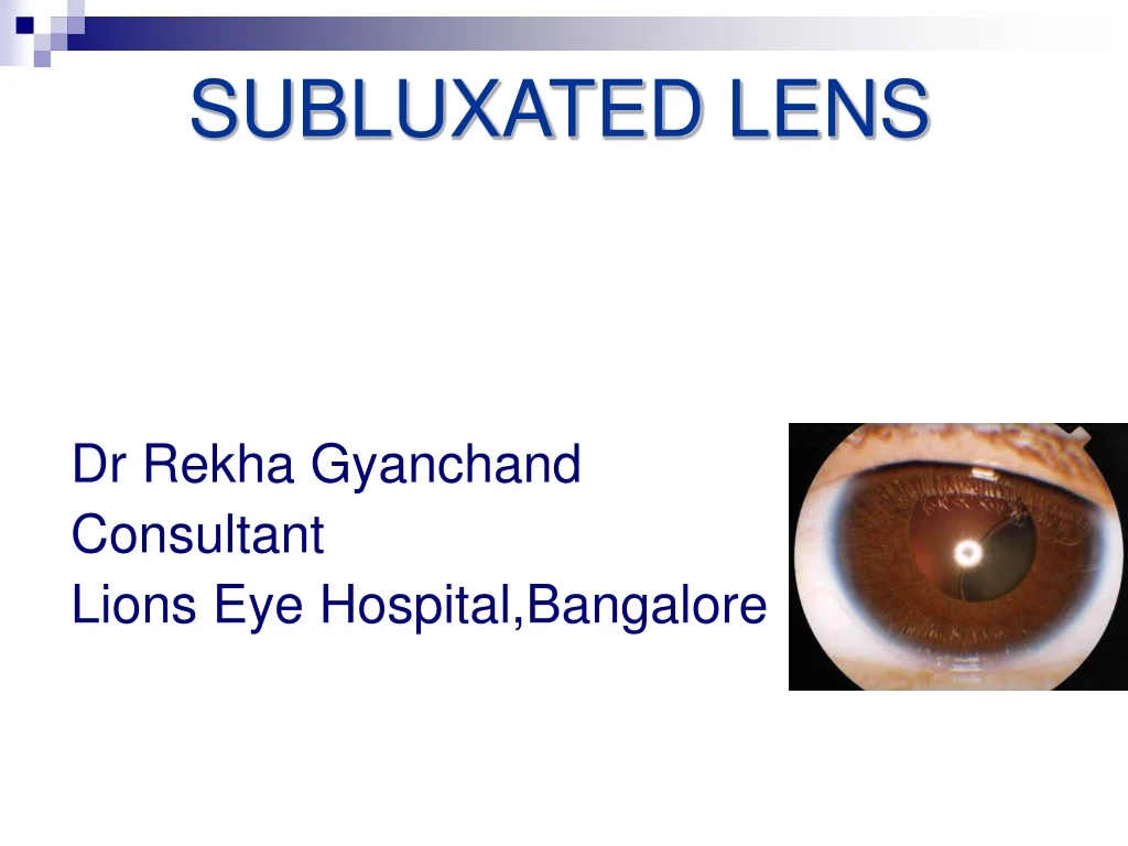
SUBLUXATED LENS. Dr Rekha Gyanchand Consultant Lions Eye Hospital,Bangalore. ANATOMY OF SUBLUXATION. The lens is suspended in its anatomic position by ciliary zonules (zonules of Zinn or suspensory ligament of Zinn)

E N D
SUBLUXATED LENS Dr Rekha Gyanchand Consultant Lions Eye Hospital,Bangalore
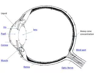
ANATOMY OF SUBLUXATION • The lens is suspended in its anatomic position by ciliary zonules (zonules of Zinn or suspensory ligament of Zinn) • Zonules fibers which run from ciliary body and fuse into the outer layer of the lens capsule around the equatorial zone. • The bundles that insert into the anterior capsule are stronger than those that insert in the posterior capsule. • The insertion of zonules extends 1.5 mm anterior to the equator and 1mm posterior . • Each zonule measures 5 to 30µm in diameter and is composed of bundles of microfibrils. • Biochemically they are composed of fibrillin, a protein product of the gene linked to Marfan’s syndrome.
Definition -Subluxation of lens Malposition of the crystalline lens when it is partially displaced but contained within the lens space .Few zonular attachments present. Dislocation of lens Lens lies completely outside the lens patellar fossa, in the anterior chamber, free-floating in the vitreous, or directly on the retina
CONGENITAL Isolated ectopia lentis Ectopia lentis et pupillae Hereditary ectopia lentis with systemic manifestations (Marfens etc) ACQUIRED Trauma-Common WEAK ZONULES Hypermature cataract Chronic cyclitis , uveitis RP ,RD Syphilis Buphthalmos Severe or pathological myopia Ciliary body tumor ETIOLOGY
Congenital -Single (isolated) ectopia lentis Autosomal dominant inheritance Birth, late onset has been described Dysfunctional zonular apparatus Superotemporal displacement
Congenital - Symmetric eccentric pupils Autosomal recessive Bilateral Irides often appear, Atrophic & Transillumination defects Dysfunctional zonular fibers Displaced in the opposite direction of the lens subluxation .
Congenital- Hereditary ectopia lentis with systemic manifestations
Marfan Syndrome- Heritable Ectopia lentis Lens subluxated - 75% bilateral, symmetrical, supertemporal Axial myopia ,Increase incidence of RD
Homocystinuria- In born metabolic error Fair skin with coarse hair Osteoporosis MR (50 %) Seizure disorder Marfanoid habitus Poor circulation Lens subluxated – 90% Bilateral, Symmetrical Inferonasal,
Weil Marchesani Syndrome Ectopia lentis Microspherophakia lenticular myopia High incidence of lens subluxation occurs inferiorly, often progressing to complete dislocation. Pupillary block glaucoma is common; therefore, prophylactic laser peripheral iridotomies are recommended.
ASSOCIATED OCULAR ANOMALIES • Congenital glaucoma/buphthalmos • Pseudoexfoliation syndrome • Chronic uveitis • Retinitis pigmentosa • Megalocornea • Aniridia • Hypermature cataract • High myopia
Others .. Sulfite oxidase deficiency Extremely rare Defect in sulfur metabolism. Progressive CNS abnormalities - within the first year of life in concert with ectopia lentis Hyperlysinemia Autosomal recessive Enzymatic defect of amino acid metabolism Mental retardation Lens dislocation. Diagnosis: increased plasma levels of lysine.
Cause for decrease VA • Fluctuating vision dramatically as the vision may alternate between phakia and aphakic • Progressive movement of the lens -Extreme hyperopic or myopic shift , astigmatism • Monocular diplopia • Poor near vision (loss of accommodative power)
COMPLICATION • Pupillary block glaucoma • Forwards displacement-Endothelial damage,sec.Ac angle closure
WORK –UP History • History of ocular trauma. • Systemic history investigating systemic disease . • Cardiovascular disease (eg, Marfan syndrome) • Skeletal problems • Marfan syndrome • Weil-Marchesani syndrome • Homocystinuria • Family history • Consanguinity • Mental retardation • Unexplained deaths at young age (eg, autosomal recessive conditions, including homocystinuria, hyperlysinemia, ectopia lentis et pupillae, or sulfite oxidase deficiency)
Ocular Examination Attention to orbital anatomy : evaluate for hereditary malformations (eg, enophthalmos with facial myopathic appearance seen in patients with Marfan syndrome). Measure corneal diameter (megalocornea is associated with Marfan syndrome). Strabismus (amblyopia). Careful retinoscopy and refraction is essential, often revealing myopia with astigmatism. Keratometry may help ascertain degree of corneal astigmatism.
Ocular Evaluation VA Near /distant BCVA, Best vision with aphakic correction Gonioscopy note any developmental defects, pseudoexfoliative material and deformities secondary to trauma or as a sequlae to subluxation. Fundus examination is done to look for lattice degeneration, cyclitic membranes, retinal detachment or posttraumatic pathology. Retinal detachments occur in 10% eyes with Marfan’s syndrome and Homocystinuria. Bscan ultrasonography is indicated in opaque ocular media. Presence if any of uveitis, glaucoma, corneal edema and amblyopia should also be ascertained Ultrasound biomicroscopy /anterior segment OCT, are especially useful for zonular and angle assessment in patients where the pupil fails to dilate A-Scan Axial length measurement Causes of glaucoma in ectopia lentis pupillary block , phacoanaphylaxis or phacolytic , posttraumatic angle recession poorly developed angle structures , lens in the anterior chamber.
Preoperative Evaluation • Evaluate lens position, and identify phacodonesis or cataract • Exact degree of zonular loss • Vitreous in the anterior chamber . • An inferior subluxation often indicates 360 degrees of zonular insufficiency combined with the effect of gravity.
OTHER INVESTIGATIONS Laboratory Studies Perform appropriate diagnostic and laboratory evaluation, if a hereditary condition is suspected - cardiac evaluation for Marfan syndrome - check serum and urine levels of homocysteine or methionine for homocystinuria). Imaging Studies Echography
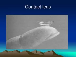
Conservative Measures • For stable induced refractive errors, visual correction with glasses or contact lenses may be an option. • Partially occluding contact lenses • Cycloplegics/miotics • Enlarge aphakic portion - Nd YAG zonulolysis - Optical iridectomy
Indication for Lensectomy Lens in the anterior chamber Lens-induced uveitis Lens-induced glaucoma Lenticular opacity with poor visual function Cataract Anisometropia or unstable refractive error Impending dislocation of the lens : large zonular dialysis
Dos and Don’ts - Cataract Incisions • Preferably away from area of zonular weakness • Use high molecular weight viscoelastic • Capsulorrhexis should be initiated in an area remote from the dialysis ,Capsulorrhexis is more easily performed with forceps than with cystitome & should be made "off-center" in an eye with significant lens subluxation
CAPSULAR TENSION RINGS • Morcher CTR • Alcon CTR • Cionni CTR • CTR Ringmorcher
Choice of surgical procedureDegree of Zonular dehicense Procedure chosen • Superior upto 4 clock hours • 1st choice: CTR with IOL implantation • 2 st choice: IOL implantation with haptic being used to stretch the bag • Inferior upto 3 clock hours CTR with IOL implantation • Anywhere> 3 to 6 clock hours Modified CTR with single loop • Anywhere >6 to >9 clock hours Modified CTR with double loop with IOL implantation • 9 or more clock hours /generalized weakness of zonulesIntracapsular cataract extraction with scleral fixated IOL/Iris fixated IOL/anterior chamber IOL
Surgical Technique Anterior limbal approach ICCE ECCE SICS PHACO Posterior pars plana approach Vitrectomy : soft lens Phacofragmentome
Surgical tips Good sized rhexis err on larger side … they develop capsule contraction Use a chop or supracapsular technique, because it allows to raise the nucleus above the capsule, putting less stress on the zonules. Another technique is viscoelevation.
Counselling Patient Education Genetic counselling Follow up with physician to rule out life-threatening disorders. Safety glasses are advocated when risk of eye injury is possible.