Download
1 / 19
210 likes | 624 Views
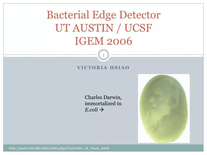
Bacterial Edge Detector UT AUSTIN / UCSF IGEM 2006. Victoria hsiao . Charles Darwin, immortalized in E.coli . Projects mentioned in the presentation. Bacterial Edge Detector – engineering E.coli to detect light/dark boundaries and make a black outline only on those edges.

E N D
Bacterial Edge DetectorUT AUSTIN / UCSF IGEM 2006 Victoria hsiao Charles Darwin, immortalized in E.coli http://parts.mit.edu/wiki/index.php/University_of_Texas_2006
Projects mentioned in the presentation • Bacterial Edge Detector – engineering E.coli to detect light/dark boundaries and make a black outline only on those edges. • Regulating chemotaxis in E.coli – engineering E.coli to “tumble” in place instead of swimming straight, thus making them stationary. • Photofabrication – using mask-directed lithography to make 3D microstructures from cross-linked proteins that living organisms can interact with. “Spatial Recognition of Bacterial Populations” UT Austin/ UCSF iGEM 2006 Presentation Ppt
Engineering a Bacterial Edge Detector iGEM 2006 UT Austin/UCSF Lawn of E.coli Printed transparency “Spatial Recognition of Bacterial Populations” UT Austin/ UCSF iGEM 2006 Presentation Ppt
Steps to Engineering a Bacterial Edge Detector • 1. Make E.coli (which is blind) able to detect light (iGEM 2005) • 2. Capture an image with a lawn of E.coli (iGEM 2005) • 3. Make the E.coli compute the light/dark boundary of the captured image (iGEM 2006) “Spatial Recognition of Bacterial Populations” UT Austin/ UCSF iGEM 2006 Presentation Ppt
Background: Bacterial Photography (2005) • 1. Getting E.coli to detect light • Make Cph1 (photoreceptor) from heme by using the genes ho1 and pcyA (derived from cyanobacteria) • Cph1 is combined with the histidine kinase Env Z (which e.coli already had) to make the chimera Cph8. Images: http://parts.mit.edu/wiki/index.php/University_of_Texas_2006
Photography: Light vs Dark In the dark, the Env Z phosphorylates the OmpR transcription factor protein, which binds to the promoter PompC, which expresses the reporter LacZ, which will produce a black precipitate when the sugar S-gal is added. When the E.coli is exposed to 660 nm light, the transcription cascade is repressed because Cph 1 isomerizes and changes its conformation, which inactivates the Env Z. Thus, these sections remain light. Images: http://parts.mit.edu/wiki/index.php/University_of_Texas_2006
Light Imaging Setup Mercury lamp 632nm bandpass filter 35mm slide Double Gauss focusable lens Projected Image “Engineering Escherichia Coli to see light” Levskaya et al. Nature, Brief Communications 2005 (Supplementary Materials)
Steps 1 & 2 Completed • Light areas are light, dark areas are dark, but can we make an outline? (2005) The E.coli could make a high-contrast replica of the projected image (2006) Step 3 : taking the projected image and only showing expressing black at the edges of light and dark “Spatial Recognition of Bacterial Populations” UT Austin/ UCSF iGEM 2006 Presentation Ppt
Edge Detection (2006) • Transcription cascade is “black-boxed” into an inverter block Red light Gene expression repressed! “Spatial Recognition of Bacterial Populations” UT Austin/ UCSF iGEM 2006 Presentation Ppt
The Edge Detection Circuitry Lux 1 gene – lacZ activator, produces AHL (acylated homoserine lactone) which binds to LuxR. C1 gene – lacZ dominant repressor, produces c1 which binds to Oλ , repressing lacZ even if lux1 is activated. Therefore, the only way to get a black output is to have AHL, but while c1 is repressed. How? Images: http://parts.mit.edu/wiki/index.php/University_of_Texas_2006
Edge Detection Logic (continued) Dark Light In both cases, light and dark, lacZ expression is repressed. However, the AHL produced by the dark bacteria is able to diffuse to surrounding bacteria, and only the light bacteria will be able to use it . Images: http://parts.mit.edu/wiki/index.php/University_of_Texas_2006
Edge Detection Logic (continued) Therefore, this case can only occur in light bacteria at the light/dark boundary, and the E.coli can detect edges.. Images: http://parts.mit.edu/wiki/index.php/University_of_Texas_2006
Leaky C1 light repression When light was projected onto the E.coli the C1 gene wasn’t being entirely repressed, so when the AHL diffused over from the dark bacteria, lacZ was still repressed. Images: http://parts.mit.edu/wiki/index.php/University_of_Texas_2006
Toning Down C1 Expression with RBS By adding ribosomal binding sites (RBS) to the gene sequence, they were able to tone down the expression of C1 such that it was still dominant in the dark, but permissive in the light. So they tried 3 different concentrations of RBS: RBS3 0.07x was the only one that worked. “Spatial Recognition of Bacterial Populations” UT Austin/ UCSF iGEM 2006 Presentation Ppt
It Worked! Images: http://parts.mit.edu/wiki/index.php/University_of_Texas_2006
Improving Contrast & Sharpness of Edge Detection • LuxI poison that is expressed in light so that there is less of a gradient at the edge. • Mix in Aiia (anti-AHL) expressing strain to take up AHL at different rates to alter the width of the edge. • Modify pH (AHL is destabilized by > 7.5) “Spatial Recognition of Bacterial Populations” UT Austin/ UCSF iGEM 2006 Presentation Ppt
Other things they found • In all the experiments just described they used a two plasmid system to transform the E.coli: • Plasmid 1 contained the phycobilins ho1 and pcyA (which make photoreceptor in Cph1) • Plasmid 2 contained Cph8 • When they combined the two plasmids into a single plasmid (Bba_M30109), they got inverted logic. So now, light activated both luxI and ch1 while dark repressed. But then they noted that the background signal of Bba_M30109 was too high for bacterial photographs. • They also found that Cph1 responds to another wavelength in addition to the 660nm. A 735nm wavelength changes the Cph1 conformation in such a way that the dark conditions are activated. This is useful because a 735nm light can sometimes be aimed more precisely than a projected shadow. “Spatial Recognition of Bacterial Populations” UT Austin/ UCSF iGEM 2006 Presentation Ppt
Things I Thought Were Exciting • E.coli can be madeinto light sensors just by combining photoreceptor genes from cyanobacteria with genes for an enzyme that E.coli already has. • Making each cell do a simple computation, so that having an entire lawn of bacteria results in massive parallel computations.
Sources • “Engineering Escherichia Coli to see light” Levskaya et al. Nature, Brief Communications 2005 (Supplementary Materials) • “Spatial Recognition of Bacterial Populations” UT Austin/ UCSF iGEM 2006 Presentation Ppt • UT Austin iGEM Wiki page, http://parts.mit.edu/wiki/index.php/University_of_Texas_2006