Download
1 / 43
520 likes | 2.33k Views
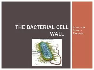
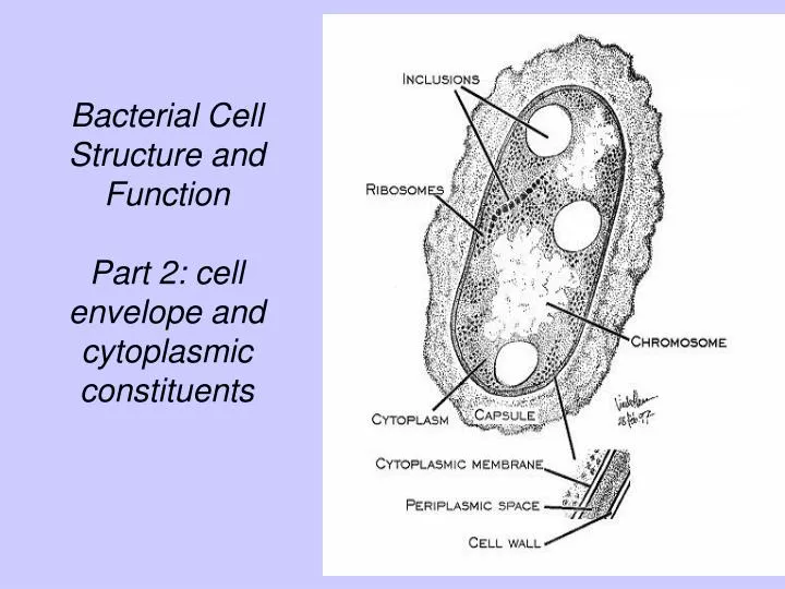
Bacterial Cell Structure and Function Part 2: cell envelope and cytoplasmic constituents. Cell Walls. Cell wall is a structure that completely surrounds the cell protoplast. (Almost) all bacteria have a cell wall. Cell Walls. Why study bacterial cell walls?

E N D
Bacterial Cell Structure andFunctionPart 2: cell envelope and cytoplasmic constituents
Cell Walls • Cell wall is a structure that completely surrounds the cell protoplast. • (Almost) all bacteria have a cell wall.
Cell Walls Why study bacterial cell walls? • They are essential structures in bacteria. • They are made of chemical components found nowhere else in nature. • They may cause symptoms of disease in animals. • They are the site of action of some of our most important antibiotics.
Profile of the bacterial cell envelope • Gram-positive cell wall is thick homogeneous monolayer • Gram-negative cell wall is thin heterogeneous multilayer
Primary function of the bacterial cell wall • To prevent rupture or osmotic lysis of the cell protoplast Lysis of a pair of dividing E. coli cells
Chemical nature of bacterial cell walls • Bacterial cell walls always contain murein, which is a type of peptidoglycan • Chemical nature of murein accounts for the function of the cell wall • Murein is only found in the cell walls of bacteria E. coli peptidoglycan
Chemical nature of bacterial cell walls Peptidoglycan is made up of • 2 amino sugars N-acetyl-glucosamine = G N- acetylmuramic acid = M • 4 amino acidsL-alanine = L-alaD-glutamic acid = D-gludiaminopimelic acid = DAPD-alanine = D-ala
Chemical nature of bacterial cell walls Gram-negative murein. Murein is a polymer of the peptidoglycan subunit. The sugars form the glycan backbone (G-M-G-M-etc.) and the amino acids comprise the peptide side chains of the molecule.
Chemical nature of bacterial cell walls Penicillin prevents formation of this Interpeptide bond Gram-negative murein showing the sites of action of the antibiotic penicillin and the enzyme lysozyme Lysozyme breaks this glycoside bond between M and G
Chemical nature of bacterial cell walls Gram-positive murein has a thicker glycan a backbone and there are interpeptide bridges that join amino acid side chains together.
Chemical nature of bacterial cell walls Gram-positive murein showing the sites of action of the antibiotic penicillin and the enzyme lysozyme Penicillin blocks the Insertion of the inter- peptide bridge Lysozyme breaks the glycoside bond between M and G
Other characteristics of bacterial cell walls Gram-positive cell walls contain teichoic acids Teichoic acids are thought to stabilize the Gram positive cell wall and may be used in adherence.
Other characteristics of bacterial cell walls Gram-negative cell walls include an outer membrane
Other characteristics of bacterial cell walls Outer membrane of Gram-negatives has two important properties • It protects the cells from permeability by many substances including penicillin and lysozyme. • It is the location of lipopolysaccharide (endotoxin) which is toxic for animals.
Table: Correlation of the Gram stain with properties of bacterial cell walls
Cell (cytoplasmic) membrane • Completely encloses the bacterial cell protoplast • Composed of 60% protein and 40% phospholipid • Arranged as a bilayer Section of a cytoplasmic membrane
Membrane structure and assembly • The membrane bilayer is formed by phospholipidmolecules made up of glycerol and fatty acids
Membrane structure and assembly • Phospholipids arrange themselves spontaneously in water: lipid “tails” inward; glycerol “heads” outward
Membrane structure and assembly The proteins associate with both sides of the membrane, or may imbed in the membrane, or pass through the membrane. The fluid mosaic model of a membrane
Membrane structure and assembly Proteins in the cytoplasmic membrane have a variety of functions including transport and energy transformations The cytoplasmic membrane of E. coli
Functions of the cytoplasmic membrane • Osmotic or permeability barrier: the membrane is impermeable to molecules that are charged or greater than molecular weight of 100
Functions of the cytoplasmic membrane • Location of transport systems to import all the needed molecules that are charged or greater than molecular weight 100
Functions of the cytoplasmic membrane • Energy generation: location of the electron transport system (ETS) and the ATP synthsizing enzyme ATPase
Functions of the cytoplasmic membrane • Specialized functions involving cell wall synthesis, cell division and DNA replication.
Cytoplasmic Constituents of Bacterial Cells • Cytoplasm • Genetic material: chromosome and Plasmids (DNA) • Ribosomes • Inclusions
Cytoplasm of bacterial cells is gel-like and contains the chromosome, ribosomes,various macromolecules and small molecules in water solution. ribosomes DNA (chromosome)
Small molecules present in a growing bacterial cell Molecules Approximate number of kinds Amino acids, their precursors and derivatives 120 Nucleotides, their precursors and derivatives 100 Fatty acids and their precursors 50 Sugars, carbohydrates and their precursors or derivatives 250 quinones, porphyrins, vitamins, coenzymes and prosthetic groups and their precursors 300 Ions (PO4, NH3, SO4, etc.) 20
The Bacterial Chromosome or “Nucleoid” Bacterial DNA released from a “gently lysed” E. coli cell
DNA (deoxyribonucleic acid) is the genetic material of the cell. It can be replicated in a semiconservative fashion and passed on to progeny cells.
Ribosome Structure and Composition The procaryotic ribosome (L) is 70S in size, being composed of a 50S (large) subunit and a and 30S (small) subunit. The eucaryotic ribosome (R) is 80S in size and is composed of a 60S and a 40S subunit.
Ribosome Structure and Composition Ribosomes are made of two subunits, a large subunit and a small subunit. Each subunit is made up of RNA and various proteins.
Ribosome Function Ribosomes function in protein synthesis. Amino acids are assembled into proteins according to the genetic code on the surfaces of ribosomes during the process of translation.
Some inclusions in Bacterial Cells Function
Some inclusions in Bacterial Cells Bacterial Inclusions. A. PHB granules; b. a parasporal BT crystal in the sporangium of Bacillus thuringiensis; c. carboxysomes in Anabaena viriabilis, showing their polyhedral shape; d. sulfur globules in the cytoplasm of Beggiatoa.
Endospores are produced as intracellular structures within the cytoplasm of certain bacteria, most notably Bacillus and Clostridium species. Endospore forming bacteria left to right: Clostridium botulinum, Bacillus brevis, Bacillus thuringiensis
Endospore formation is NOT a mechanism of reproduction. Rather it is a mechanism for survival in deleterious environments. During the process of spore formation, one vegetative cell develops into one endospore. The sequential steps of endospore formation in a Bacillus species. The process of endospore formation takes about six hours. Eventually the mature endospore is released from its “mother cell” as a free spore Free endospore Endospore within mother cell Vegetative cell
Under favorable nutritional and environmental conditions, an endospore germinates into a vegetative cell.
Medically-important Endospore-forming Bacteria • Bacillus anthracis causes anthrax • Bacillus cereus causes food poisoning • Clostridium tetani causes tetanus • Clostridium botulinum causes botulism • Clostridium perfringens causes food poisoning and gas gangrene • Clostridium difficile causes antibiotic-induced diarrhea and pseudomembranous colitis
Properties of Endospores • Resting (dormant) cells - “cryptobiotic” i.e., show no signs of life…..primarily due to lack of water in the spore
Properties of Endospores • Several unique surface layers not found in vegetative cells: exosporium, spore coat, cortex, and core wall
Properties of Endospores • Highly resistant to heat (boiling), acids, bases, dyes ( don’t stain) irradiation, disinfectants, antibiotics, etc.
Properties of Endospores • Spores and parasporal crystals produced by some bacteria are toxic to insects Parasporal crystal Endospore