Download
1 / 40
741 likes | 2.77k Views
Osteoarthritis. Osteoarthritis (OA). OA is the most common form of arthritis and the most common joint disease Over 10 million Americans suffer from OA of the knee alone Most of the people who have OA are older than age 45, and women are more commonly affected than men.

E N D
Osteoarthritis (OA) • OA is the most common form of arthritis and the most common joint disease • Over 10 million Americans suffer from OA of the knee alone • Most of the people who have OA are older than age 45, and women are more commonly affected than men. • OA most often occurs at the ends of the fingers, thumbs, neck, lower back, knees, and hips.
OA • OA is a disease of joints that affects all of the weight-bearing components of the joint: • Articular cartilage • Menisci • Bone
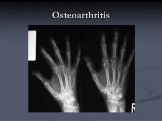
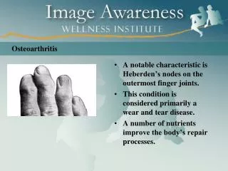
OA Nodal osteoarthritis Note bony enlargement of distal and proximal interphalangeal joints (Heberden's nodes and Bouchard's nodes, respectively).
OA – Risk Factors Age • Age is the strongest risk factor for OA. Although OA can start in young adulthood, if you are over 45 years old, you are at higher risk. Female gender • In general, arthritis occurs more frequently in women than in men. Before age 45, OA occurs more frequently in men; after age 45, OA is more common in women. OA of the hand is particularly common among women. Joint alignment • People with joints that move or fit together incorrectly, such as bow legs, a dislocated hip, or double-jointedness, are more likely to develop OA in those joints.
OA – Risk Factors Hereditary gene defect • A defect in one of thegenes responsible for the cartilage component collagen can cause deterioration of cartilage. Joint injury or overuse caused by physical labor or sports • Traumatic injury (ex. Ligament or meniscal tears) to the knee or hip increases your risk for developing OA in these joints. Joints that are used repeatedly in certain jobs may be more likely to develop OA because of injury or overuse. Obesity • Being overweight during midlife or the later years is among the strongest risk factors for OA of the knee.
OA – Symptoms • OA usually occurs slowly - It may be many years before the damage to the joint becomes noticeable • Only a third of people whose X-rays show OA report pain or other symptoms: • Steady or intermittent pain in a joint • Stiffness that tends to follow periods of inactivity, such as sleep or sitting • Swelling or tenderness in one or more joints [not necessarily occurring on both sides of the body at the same time] • Crunching feeling or sound of bone rubbing on bone (called crepitus) when the joint is used
Osteoarthritis (OA) - Definition Osteoarthritis may result from wear and tear on the joint • The normal cartilage lining is gradually worn away and the underlying bone is exposed.
The repair mechanisms of tissue absorption and synthesis get out of balance and result in osteophyte formation (bone spurs) and bone cysts Osteoarthritis (OA) - Definition A case of the, “Which came first? The chicken or the egg?”
OA – Articular Cartilage • Articular cartilage is the main tissue affected • OA results in: • Increased tissue swelling • Change in color • Cartilage fibrillation • Cartilage erosion down to subchondral bone
OA – Articular Cartilage A) Normal articular cartilage from 21-year old adult (3000X) B) Osteoarthritic cartilage (3000X) The surface changes alter the distribution of biomechanical forces further triggering active changes by the tissue
OA – Articular Cartilage The cartilage damage causes chondrocyte cloning in an attempt to restore articular surface (Normal adult chondrocytes are fully differentiated and do not proliferate) (A) Normal articular cartilage (B) Osteoarthritic cartilage
OA – Articular Cartilage • Unfortunately, the newly dividing cells do not differentiate fully and cannot effectively synthesize the elements needed for matrix maintenance • This results in a net loss of matrix components • Collagen content stays constant but fibrils are thinner and more disorganized • - Decreased tensile strength
OA – Articular Cartilage • Proteoglycan loss results in an inability to hold on to water content: • - Decreased resistance to compression – especially with repeated stress
OA vs. Aging Unlike aging, OA is progressive and a significantly more active process
OA – Overall Changes Osteoarthritis with lateral osteophyte, loss of articular cartilage and some subchondral bony sclerosis- X-ray shows loss of joint space
OA – Radiographic Diagnosis Asymmetrical joint space narrowing from loss of articular cartilage The medial (inside) part of the knee is most commonly affected by osteoarthritis.
OA – Radiographic Diagnosis • Asymmetrical joint space narrowing • Periarticular sclerosis • Osteophytes • Sub-chrondral bone cysts
Normal Articular Cartilage Ostearthritic degenerated cartilage with exposed subchondral bone OA – Arthroscopic Diagnosis Arthroscopy allows earlier diagnosis by demonstrating the more subtle cartilage changes that are not visible on x-ray
OA – Arthroscopic Treatment • In addition to being the most accurate way of determining how advanced the osteoarthritis is: • Arthroscopy also allows the surgeon to debride the knee joint • Debridement essentially consists of cleaning out the joint of all debris and loose fragments. During the debridment any loose fragments of cartilage are removed and the knee is washed with a saline solution. • The areas of the knee joint which are badly worn may be roughened with a burr to promote the growth of new cartilage - a fibrocartilage material that is similar scar tissue. • Debridement of the knee using the arthroscope is not 100% successful. If successful, it usually affords temporary relief of symptoms for somewhere between 6 months - 2 years. • Arthroscopy also allows access for surgical treatment of articular cartilage: graft-transplantation, micro-fracture techniques, sub-chondral drilling
OA – Disease Management • OA is a condition which progresses slowly over a period of many years and cannot be cured • Treatment is directed at decreasing the symptoms of the condition, and slowing the progress of the condition • Functional treatment goals: • Limit pain • Increase range of motion • Increase muscle strength
OA – Non-operative Treatments • Pain medications • Physical therapy • Walking aids • Shock absorption • Re-alignment through orthotics • Limit strain to affected areas
Proximal Tibial Osteotomy • Osteoarthritis usually affects the inside half (medial compartment) of the knee more often than the outside (lateral compartment). • This can lead to the lower extremity becoming slightly bowlegged, or in medical terms, a genu varum deformity
Proximal Tibial Osteotomy • The result is that the weight bearing line of the lower extremity moves more medially (towards the medial compartment of the knee). • The end result is that there is more pressure on the medial joint surfaces, which leads to more pain and faster degeneration. • In some cases, re-aligning the angles in the lower extremity can result in shifting the weight-bearing line to the lateral compartment of the knee. This, presumably, places the majority of the weight-bearing force into a healthier compartment. The result is to reduce the pain and delay the progression of the degeneration of the medial compartment.
Proximal Tibial Osteotomy • In the procedure to realign the angles, a wedge of bone is removed from the lateral side of the upper tibia. • A staple or plate and screws are used to hold the bone in place until it heals. • This converts the extremity from being bow-legged to knock-kneed. • The Proximal Tibial Osteotomy buys some time before ultimately needing to perform a total knee replacement. The operation probably lasts for 5-7 years if successful.
Total Knee Replacement • The ultimate solution for osteoarthritis of the knee is to replace the joint surfaces with an artificial knee joint: • Usually only considered in people over the age of 60 • Artificial knee joints last about 12 years in an elderly population • Not recommended in younger patients because: • The younger the patient, the more likely the artificial joint will fail • Replacing the knee the second and third time is much harder and much less likely to succeed. • Younger patients are more active and place more stress on the artificial joint, that can lead to loosening and failure earlier • Younger patients are also more likely to outlive their artificial joint, and will almost surely require a revision at some point down the road. • Younger patients sometimes require the surgery (simply because no other acceptable solution is available to treat their condition)
Total Knee Replacement • The ends of the femur, tibia, and patella are shaped to accept the artificial surfaces. • The end result is that all moving surfaces of the knee are metal against plastic
Total Knee Replacement Photographs of total knee components on model bone
Unicompartmental Knee Replacement • When only one part of the knee joint is arthritic, it may be possible to replace just this part of the joint • The procedure is similar to a total knee replacement, but only one side of the joint is resurfaced • A metal component is fit onto the femur and a plastic bearing is inserted either directly onto the tibia or onto a metal tray which has been fit onto the tibia • Recovery time is generally slightly shorter following this kind of surgery.