Download
1 / 28
280 likes | 852 Views
discolored skin caused by benign tumors of dermal blood capillaries (strawberry ... Skin cancer. induced by UV rays of the sun. most common in fair-skinned ...

E N D
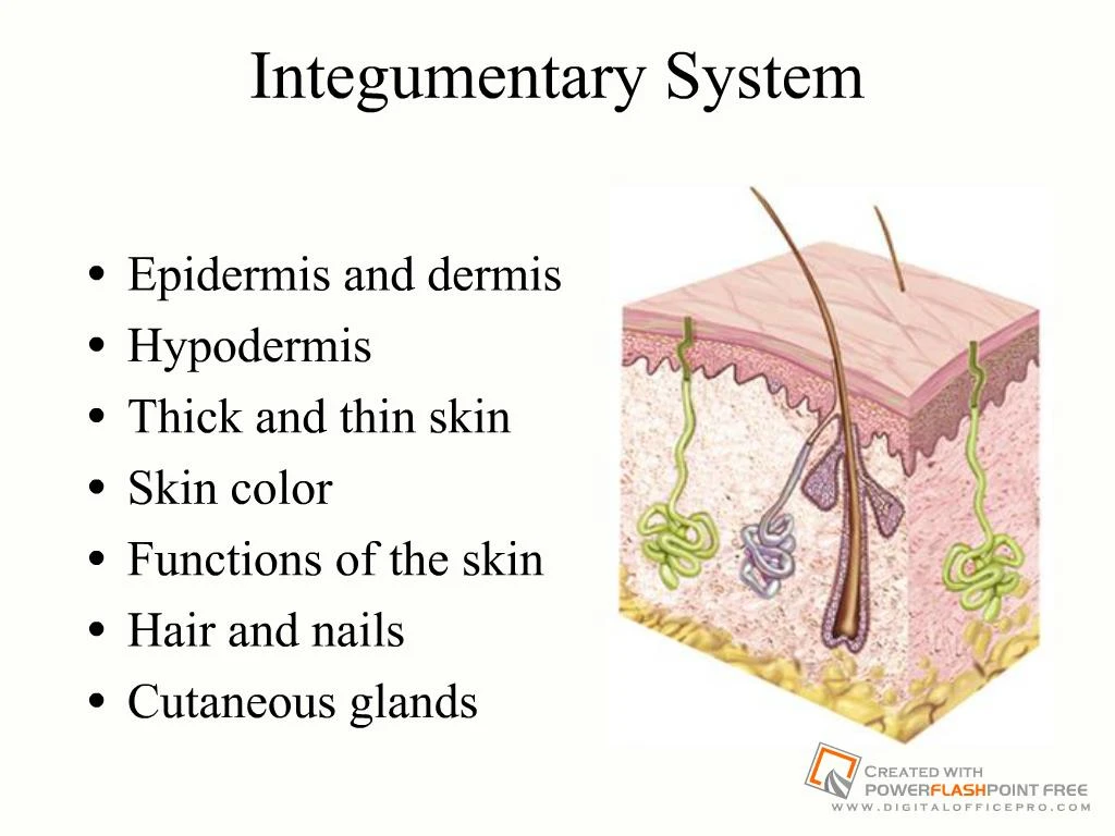
Slide 1:Integumentary System
Epidermis and dermis Hypodermis Thick and thin skin Skin color Functions of the skin Hair and nails Cutaneous glands
Slide 2:Overview of the Skin
Largest organ of the body (16% of body weight) Two layers epidermis keratinized stratified squamous epithelium contains ______________ dermis connective tissue layer Rests on subcutaneous layer or hypodermis Normal thickness of 0.5 mm, up to 5 mm
Slide 3:Cell Types & Layers of the Epidermis
Slide 4:Dermis
Thicker than epidermis Composition collagen, elastic & reticular fibers, fibroblasts & accessory structures such as hair follicles and glands Dermal papillae Layers papillary layer is areolar tissue & dermal papillae of upper dermis ______________ layer is deeper part of dermis
Slide 5:Layers of the Dermis
Slide 6:Hypodermis
Known as subcutaneous tissue or superficial fascia Has more adipose than dermis Functions energy reservoir ______________________ Hypodermis
Slide 7:Skin Colors (Pigmentation)
________________ is red pigment of red blood cells visible through dermal collagen fibers ________ is yellow pigment of vegetables & egg yolks concentrates in stratum corneum & subcutaneous fat Melanin pigment produced by ___________________ pigment synthesis stimulated by UV radiation from sunlight produces yellow, brown, black and reddish hues
Slide 8:Abnormal Skin Colors
Cyanosis - blueness from lack of oxygen Erythema - redness from dilated cutaneous vessels Jaundice - yellowing of skin & sclera - bilirubin Bronzing - golden-brown color of Addison disease (deficiency of glucocorticoid hormone) Pallor - pale color from lack of blood flow Albinism - a genetic lack of melanin Hematoma - a bruise (visible clotted blood)
Slide 9:Skin Markings
Hemangiomas (birthmarks) discolored skin caused by benign tumors of dermal blood capillaries (strawberry birthmarks disappear in childhood -- port wine birthmarks last for life) Freckles & moles = aggregations of melanocytes freckles are flat; moles are elevated Friction (epidermal) ridges leave oily fingerprints
Slide 10:Functions of the Skin
Barrier = tough, dry, acid mantle, water barrier, UV barrier Vitamin D synthesis UV light converts 7-dehydrocholesterol in dermal vessels to vitamin D3 Cutaneous absorption 1-2 % oxygen absorption by diffusion through skin fat-soluble vitamins (A, D, E & K) easily absorbed Sensory functions receptors for heat, cold, touch, pressure, vibration & pain Thermoregulation cutaneous vasodilation & constriction and sweating Psychological and social functions appearance & social acceptance facial expression and nonverbal communication
Slide 11:Characteristics of Human Hair
S. corneum of the skin contains soft keratin Hair and nails are composed of hard keratin toughened by disulfide bridges between molecules Hair found almost everywhere on the body 3 different body hair types lanugo -- fine, unpigmented fetal hair vellus -- fine, unpigmented hair terminal hair -- coarse, long, pigmented hair
Slide 12:Structure of Hair and Follicle
Hair is filament of keratinized cells Shaft: parts above skin Root: parts below within follicle Follicle: epidermal invagination into dermis Cross section layers: medulla, cortex and cuticle Bulb: swelling in base where hair originates Papilla: vascular tissue in bulb Hair color is due to melanin pigments eumelanin pheomelanin (agouti signaling protein)
Slide 13:Structure of Hair Follicle
Epithelial root sheath is an extension of the epidermis Connective tissue root sheath is derived from the dermis Hair receptors entwine each follicle Arrector pili muscle
Slide 14:Growth of Hair
Mitosis in stratum basale of epithelial root sheath as cells become keratinized they are pushed upward Grow 1 mm every 3 days for 2 to 5 years dormant phase lasts several months to years as new hair begins to grow it pushes out old hair eyelashes and eyebrows only grow for about a month then are dormant for 3 months
Slide 15:Functions of Hair
Body hair too thin to provide warmth Sensory functions alert us to parasites crawling on skin Scalp hair provides heat retention & sunburn cover Sex and individual recognition Beard, pubic & axillary hair indicate sexual maturity & help distribute sexual scents Guard hairs & eyelashes prevent foreign objects from getting into nostrils, ear canals or eyes Expression of emotions with eyebrows
Slide 16:Nails
Clear, hard derivative of stratum corneum densely packed cells filled with hard keratin Flat nails allow for fleshy, sensitive fingertips Growth rate is 1 mm per week new cells added by mitosis in the nail matrix nail plate is visible part of nail
Slide 18:Cutaneous Glands
Sweat glands _______________ _______________ Sebaceous glands Ceruminous glands Mammary glands
Slide 19:Sweat Glands
Filtrate of plasma 500 ml of perspiration/day Merocrine gland - simple tubular gland millions of them cool the body Apocrine glands found only near hair follicles & respond to stress & sex body odor produced by _______________ action on fatty acids
Slide 20:Sebaceous Glands
Oily secretion called ______________ that contains broken-down cells Flask-shaped gland with duct that may open into hair follicle
Slide 21:Ceruminous Glands
Modified sweat gland Found only in external ear canal Their secretion combines with sebum to produce earwax waterproof keeps eardrum flexible
Slide 22:Breasts and Mammary Glands
Breasts of both sexes rarely contain glands Secondary sexual characteristic of females Glandular tissue - only during lactation and pregnancy modified apocrine sweat gland Mammary ridges or milk lines 2 rows of mammary glands in most mammals _____________________
Slide 23:Diseases of the Skin
Most vulnerable organ to injury & disease skin diseases common in old age Skin cancer induced by UV rays of the sun most common in fair-skinned and elderly basal cell carcinoma arises from cells of the stratum basale treated by surgical removal & radiation? squamous cell carcinoma arises from keratinocytes in the stratum spinosum if neglected, metastasis to the lymph nodes can be lethal malignant melanoma (most deadly cancer) arises from melanocytes of a preexisting mole ABCDE--asymmetry, border irregular, color mixed, diameter over 6 mm, & evolving
Slide 24:Burns
Causes of burns -- hot water, sunlight, radiation, electric shock or acids and bases Causes of deaths fluid loss, infection, & effects of dead tissue Degrees of burns 1st-degree = only the epidermis (red, painful & edema) 2nd-degree = epidermis & part of dermis (blistered) epidermis regenerates from hair follicles & sweat glands 3rd-degree = epidermis, dermis & more is destroyed often requires grafts & fibrosis may occur Treatment -- fluid replacement & infection control
Slide 25:Wound Healing of a Laceration
Damaged vessels leak blood Damaged cells & mast cells leak histamine dilates blood vessels increases blood flow increases capillary permeability Plasma seeps into wound carrying antibodies, clotting factors & WBCs
Slide 26:Wound Healing of a Laceration
Clot forms Scab forms on surface Macrophages start to clean up debris
Slide 27:Wound Healing of a Laceration
New capillaries grow Fibroblasts deposit new collagen Fibroblastic phase begins in 3-4 days & lasts up to 2 weeks Formation of granulation tissue.
Slide 28:Wound Healing of a Laceration
Surface epithelial cells multiply & spread beneath scab Scab falls off Epithelium regenerates Connective tissue forms only scar tissue (fibrosis) Remodeling phase may last 2 years Epithelial regeneration & connective tissue fibrosis.


