Download
1 / 54
650 likes | 1.62k Views
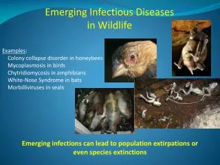
Emerging Diseases In Wildlife: Public Health and Climate Change Implications. Patrice N. Klein, MS, VMD, DACPV, DACVPM USDA APHIS Veterinary Services. Emerging Wildlife Diseases Wildlife as sentinels for global environmental alterations and climate change.

E N D
Emerging Diseases In Wildlife: Public Health and Climate Change Implications Patrice N. Klein, MS, VMD, DACPV, DACVPM USDA APHIS Veterinary Services
Emerging Wildlife DiseasesWildlife as sentinels for global environmental alterations and climate change Factors favoring disease emergence • New pathogen or more virulent existing pathogen • New host population or increase in host susceptibility • Extension in host-pathogen range • Increase in human/domestic animal/wildlife interface • Global travel (animals and humans) and trade • Antimicrobial resistance of pathogens • Habitat alteration or destruction • Climatic conditions change • Environmental contamination affects
Epidemiologic Triad The agent, the host, and the environment are always changing
Agent Host Environment Epidemiologic Triad Virulence Antibiotic susceptibility Immune reaction Distribution Vectors Survival characteristics Host range Diet Immuno-competence Exposure status Age Concurrent disease Activities Climate Demographics Mgmt. practices Culture Exposure level Stress Habitat disruption Changes in These Elements Can Cause Disease
Anthrax (bison) Adenovirus in Long-tailed ducks Avian vacuolar myelinopathy Botulism Coccidiomycosis (sea otters) Chytridiomycosis in amphibians Chronic Wasting Disease Ehrlichiosis Epizootic Hemorrhagic Disease (deer) Harmful algal blooms Hantavirus Lead toxicity Leyogonimus polyoon infection (Coots) Lyme disease Monkey-pox in Prairie dogs Morbillivirus in cetaceans Mycoplasmosis in finches Myxozoan parasite (ducks) Newcastle disease (cormorants) Plague (black-footed ferrets) Salmonellosis in redpolls West Nile Virus Toxoplasmosis (sea otters) Tularemia in Prairie dogs White-Nose syndrome (bats) “Emerging” Wildlife Diseases
Harmful Algal Blooms Toxic algae: Cylindrospermopsis Microcystis Gymnodinium breve Clinical signs: Paresis, paralysis, liver necrosis, dypsnea, diarrhea, skin rashes, eye irritation, CNS dysfunction Hawaiian Monk Seal
Harmful Algal Blooms • Brevetoxin (red tide shellfish neurotoxin); manatees and alligators (FL), Common murres (CA) • Domoic acid (amnesic shellfish toxin); California brown pelicans, cormorants, sea lions, whales (ME) • Okadaic acid (diarrheic shellfish toxin); Sea turtle fibropapilloma • Saxitoxin(paralytic shellfish toxin) • Ciguatoxin (ciguatera fish toxin); Hawaiian monk seals • Microcystin- Great Blue Herons (MD)
Epizootic Hemorrhagic Disease (EHD) • Orbiviruses • EHD (serotypes 1 and 2) • Bluetongue/BT (22 serotypes) • WTD, mule deer, pronghorn antelope, big horn sheep and cattle susceptible to both • Elk only susceptible to BT and develop diffuse hemorrhagic lesions; cattle are the BT reservoir • Transmission • Vector: Biting midges (Culicoides spp) • 10-20 days after blood meal from viremic animal, can infect new host. • Outbreaks in late summer, early fall
Epizootic Hemorrhagic Disease (EHD) • Acute form • Severe edema of the head, neck, tongue, lungs • Fever, respiratory distress • Cyanotic mucous membranes • Rapid death • Sub-acute form • Less severe than acute • Lameness, ataxia • Secondary infections • Chronic form • Fever, mild cyanosis, recovery • Sequelae- laminitis, stomatitis, rumenitis, pneumonia • Sloughed hooves, deformed coronary band growth
Epizootic Hemorrhagic Disease (EHD) • Diagnosis • Virus isolation (spleen, lymph nodes, whole blood) • Serology for chronic cases • Prevention and control • Vector control • Deer density? • Bluetongue vaccine in cattle /sheep, may cause disease in deer • Not practical • Recent outbreaks (2007, 2008) • EHD: NY, NJ, PA, OH, ID, IL, MI, WY in WTD/ pronghorn • BT: Montana in WTD and pronghorn • Expansion to Northern Latitudes???
White Nose Syndrome • Index case – NY 2007 • CT, MA, NY, VT, (PA) • Bat species affected • Little brown bat • Northern long earred bat • Small-footed bat • Eastern pipistrelle bat • Indiana bat
White Nose Syndrome • Clinical findings • Emaciation • Bats clustered in unusual locations • Flying outside hibernacula • Pathology • External fungal growth on skin, face, nose • Fungal hyphae in skin sebaceous glands • Inflammation in lungs (non-specific?) • Cause(s) – Unknown • Environment (climate change?) • Toxins? • Infectious pathogens?
Chytridiomycosis • Etiology: Batrachochytrium dendrobatidis • Distribution: • Ubiquitous in many aquatic habitats in high altitude environments • Amphibian deaths in Australia, C.A., S.A., USA. • Clinical signs: Abnormal posture, anorexia, lethargy, abnormal epidermal sloughing, ventral edema, death
Chytridiomycosis • Pathogenesis: • Fungal invasion of keratinized epidermis of adults • Alters cutaneous respiration and osmo-regulation causing death. • Healthy tadpoles are common carriers. • Diagnosis: Histopathology (affected skin) • Treatment: Antifungals (oral, topical); benzalkonium chloride or copper sulfate baths.
Trematodes (Flukes) American Coots • Etiology: Leyogonimus polyoon (introduced species) • Transmission: Indirect parasite life cycle Eggs passed in feces miracediae enter snail intermediate host • ingested by waterbirds (moorhens, coots, dabbling ducks) • gastrointestinal necrosis and death • Clinical Signs: Body weight loss, weakness, death • Diagnosis: Fecal parasitology, gross necropsy, HP • Control: Interrupt life cycle- control aquatic snails
Toxoplasmosis • Toxoplasma gondii – protozoa • Felids are definitive host • Public health • 3rd leading cause of death in foodborne illness • Pregnant women and fetal infections • Toxoplasma in marine mammals • Beluga whales, dolphins, • Sea lions, seals • California sea otters
Toxoplasmosis – CA Sea Otters • 42% Antibody positive T. gondii • 17% Deaths attributed to protozoal encephalitis • IHC / tachyzoites in brain tissue • PCR /T. gondii DNA in brain tissue • Source of fecal runoff • Feral/outdoor cats • Sewage treatment plants • Storm drain runoff • Source of infection? • Mussels, Northern anchovies
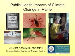
West Nile Virus Transmission Cycle Mosquito vector Incidental infections WNV WNV Incidental infections Bird reservoir hosts
West Nile Virus 2003 WILD BIRDS HUMANS
West Nile Virus 2008 Wild Birds USGS/NWHC Human USGS/CDC
West Nile Virus • Etiology: Arthropod-borne viruses/ARBOVIRUS, Flavivirus • Vectors: Culex pipiens (night feeder) and Aedes aegypti (day feeder) mosquitoes; Argasid (soft) and amblyommine (hard) ticks. Possible bird-to-bird direct transmission (experimental). • Index cases: Uganda, 1937; USA, 1999. Endemic in Africa, Middle East, Western and Central Asia. • Susceptible species: Corvids (American and fish crows, blue jays) and many avian species; Horses and people considered dead-end hosts. • Other reported species: Raccoon, skunk, rabbit, squirrel, chipmunk, bat, cat, alligators, seals, and…………
West Nile Virus • Clinical signs: Fever, ataxia, paresis, paralysis, death. Flu-like symptoms or fatal neurologic disease in people. • Gross necropsy: Severe meningeal, brain congestion; myocardial hemorrhages and necrosis; hepatic, splenic and gastrointestinal necrosis. • Histopathology: Severe vascular congestion, hemorrhage and lymphoplasmacytic inflammation in multiple tissues. • Prevention and Control: • Insect (mosquito) repellents; larvacides, adulticides. • Vaccines- Equine (killed virus)
Anthrax Incubation 1-14 days
Anthrax • Etiology: Bacillus anthracis • Anaerobic, spore-forming bacteria • Worldwide distribution; spores live in soil for years. • Recent outbreaks TX, 2005, 2007 (deer, cattle); MN, NE, ND, SD, NM 2000 (cattle, bison, horses) • All mammals are susceptible. • Ruminants (cattle, sheep, goats, bison, deer, antelope, camel) are most susceptible. • Horses, swine, dogs, cats, and humans have moderate susceptibility. • Many carnivores have natural resistance.
Anthrax • Transmission: Ingestion of contaminated water, soil, food; inhalation of spores in dust; insect bites/ skin wounds; Spores germinate in lymph nodes, multiply, and release toxins. • Clinical signs: High fever, muscle tremors, swollen lymph nodes, dysphagia, dyspnea, convulsions, colic, enteritis, bloody discharges (unclotted), death without rigor mortis. • Ataxia, sudden death, rapid bloating, bloody discharges • DO NOT OPEN CARCASS!
Anthrax • Diagnosis • Culture of blood, tissues, skin lesions • Serology (antibody titers) • Treatment: Ciprofloxacin (enrofloxacin), penicillin, tetracycline (doxycycline). • Vaccination: available for livestock and humans • Disposal: carcasses, bedding, manure – burned with wood or gasoline to cleanse the ground area
Botulism • Etiology: Clostridium botulinum toxins • Incubation period: 12-48 hrs post ingestion • Clinical Signs: Progressive paralysis of muscles- inability to fly or walk, “limber neck”, paralysis of 3rd eyelid, affects both skeletal and cardiac muscles. • Outbreaks • Multiple years: Salton Sea, CA • 2007: Lake Ontario, NY (type E) • 2008: Greak Lakes, MI (type E)
Transmission: Bacteria grow in decaying organic matter and produce toxin; maggots feed and concentrate toxin; birds ingest maggots and toxin; most common in warm months (favors bacterial growth)
Botulism • Lesions: No gross lesions • Diagnosis: Demonstration of toxin in mouse blood by serum inoculation (bioassay) • DDx: Lead poisoning, algal poisoning (toxic algal blooms), OP/carbamate pesticide poisoning, AVM • Control: Habitat management (aeration); disposal of contaminated carcasses and decaying matter. • Public Health Risk: Human cases usually Type A or B toxin (home-canned foods); Types C and E rare.
Sylvatic Plague • Etiology: Yersinia pestis • Gram negative aerobic coccobacillus • Survival briefly in soil; soft tissue (~1 wk); frozen (years) • Produces endotoxins, exotoxins, coagulase, pesticin • Enzootic • Plague maintained at steady level in rodent populations • Low death rates • Mice, voles
Sylvatic Plague • Epizootic • Large die-offs, fleas change hosts • Amplifying hosts: prairie dogs, ground squirrels, rock squirrels, wood rats, chipmunks • Black footed ferrets – outbreak 2008 • Conata Basin, South Dakota • Oral bait vaccines used on prairie dogs and BFF • Public Health • Bubonic, septicemic, pneumonic forms • Fever, lethargy,swollenLN (bubo), anorexia, death
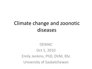
Animal Sources of Human Infection in U.S. (319 cases) 1970 -1993 Sylvatic Plague – Animal Reservoirs
Tularemia (Rabbit Fever) • Etiology - Francisella tularensis • tularensis (type A)- rabbits, squirrels • holarctica (type B)- aquatic animals • Survives mud, water, dead animals • Vector-borne transmission • Ticks (Dermacentor, Amblyomma) • Flies (Deer- fly)
Tularemia • Disease in Animals • Dogs and cats- fever, abscess at site of infection • Horses- fever, depression, stiffness • Young swine- fever depression, dyspnea • Wildlife- moribund or dead • Public health • Ulceroglandular, Typhoidal, Pneumonic • Martha’s Vineyard cases (1978, 2000)
Tularemia • Diagnosis- serology, culture, FA • Treatment- • Aminoglycosides (streptomycin, gentamicin) • Tetracyclines, cephalosporins • Prevention and Control • Insect repellents (ticks, flies) • PPE for skinning game animals • Cook game meat (rabbit, rodent) thoroughly • Avoid contaminated water (swimming and drinking)
Newcastle Disease • Etiology: Avian Paramyxovirus type 1 (APMV-1) • Transmission: contaminated feed, water, soil; aerosol (airborne); importation of exotic birds • DDx: Avian influenza Other paramyxoviruses Avian cholera Duck plague Botulism
Newcastle Disease • Exotic Newcastle Disease (END, Virulent ND) • Weakness, respiratory distress, diarrhea, periorbital edema, sudden death • Southern CA 2003 outbreak in commercial poultry • Neurotropic Virulent Newcastle Disease • Sudden severe respiratory distress, CNS signs, death • 1992-2008 sporadic outbreaks in wild Cormorants in Great Lakes region • Mesogenic: Severe pneumonia, rare CNS signs • Lentogenic: mild to severe respiratory disease
Newcastle Disease • END: Hemorrhage and necrosis of GI mucosa • Meso/Lento: Congestion in trachea and lungs; air sacculitis; Secondary bacterial respiratory infection • N.V.N.D: No gross lesions • Diagnosis: • Serology (HI, ELISA), • VI, PCR • HP
Newcastle Disease • Control: Import quarantine policies for exotic birds; disposal of infected carcasses; depopulation of infected birds; disinfection of equipment • Public Health Risk: conjunctivitis occasionally reported in people
Chronic Wasting Disease • Etiology: Abnormal prion protein (PrPcwd) Induces conformational changes in other normal prions (PrPc) over a long incubation period (years). • Clinical Signs: Behavioral changes, emaciation, weakness, ataxia, salivation, aspiration pneumonia, progressive death. • Transmission: • Saliva, feces, urine • Environmental contamination • Minimum incubation period 16 months (experimental 6 mos.) • No link to human disease thus far.
Chronic Wasting Disease • Index case: Captive mule deer in 1960’s. • Affected species: Mule deer, WTD, elk, & moose! • States: CO, WY, SD, NE, WI, NM, KS, IL, NY, WV • Recently identified in Michigan (captive WTD herd) • Samples: RPLN biopsy (deer only); CNS (obex region): rectal biopsy (elk) • Diagnosis HP and IHC
CWD Diagnosis • Immunohistochemistry (IHC) • Histopathology • Rapid ELISA • Rectal biopsy • Other tests on the horizon…..