Download
1 / 59
600 likes | 1.06k Views
GI Focused Assessment Health History. Current GI SymptomsPrevious GI ProblemsFamily History of GI ProblemsMedication Use: prescription and OTCDiet and Nutrition (Food Allergies)Use of Alcohol, street drugs, CaffeineBowel Elimination PatternSocial\Cultural Factors. GI Focused Assessment Physical .

E N D
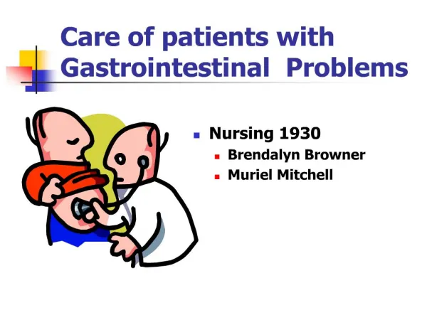
1. Care of patients with Gastrointestinal Problems Nursing 1930
Brendalyn Browner
Muriel Mitchell
3. GI Focused Assessment Physical
Vital Signs
Height and Weight
Lab and diagnostic test results
Emesis ,amount, color, consistency
Stool,amount, color, consistency, odor.
Oral Assessment
Abdominal Assessment
Rectal Assessment
4. COMMON �GI OFFENDERS� Caffeine (coffee, tea, cola)
Dairy products
Chocolate
Pepper (black and green)
Alcohol
Spicy foods
Tobacco
Drugs
5. Abdominal Assessment Inspection
Auscultation
Diaphragm (Bowel sounds)
Bell (Vascular sounds, bruits)
Percussion
Palpation
6. GI Charting Exercise
Document an assessment of the mouth in a person with normal findings.
7. EFFECTS OF AGINGPhysiologic Changes in the GI Tract Mouth
Teeth loosen, reduced circulation to gums, teeth darken and fracture
Decreased output of salivary glands
Decreased stimulation of taste buds
Stomach
Atrophy of gastric mucosa
Decreased secretion of hydrochloric acid
Decreased bile secretion
Decreased muscle tone and strength
13. Common Causes of Bleeding in the GI Tract Esophagus
Inflammation (esophagitis)
Tear (Mallory-Weiss syndrome)
Cancer
Stomach
Ulcers
Inflammation (gastritis)
Cancer
Small Intestines
Duodenal ulcer
Inflammation (Crohn�s disease)
Large Intestines and Rectum
Hemorrhoids, infections, inflammation (ulcerative colitis)
Colorectal polyps, colorectal cancer
Diverticular disease
14. GastroesophagealREFLUX DISEASE (GERD) Physiological Contributing Factors:
Incompetent lower esophageal sphincter
Irritant effects of reflux
Abnormal esophageal clearance
Delayed gastric emptying
15. GastroesophagealREFLUX DISEASE (GERD) Common Signs and Symptoms:
Heartburn
Regurgitation
Retrosternal Burning Pain (epigastrium, neck, throat)
16. GastroesophagealREFLUX DISEASE (GERD) Management and Treatment:
Lifestyle modification measures
Antacids, H2 antagonists, proton-pump inhibitor, carafate, prokinetic agents (reglan)
Surgical Intervention
Nissen Fundoplication
17. GastroesophagealREFLUX DISEASE (GERD) Pharmacology
Antacids
H2 Antagonist
Proton Pump Inhibitors (Bid)
Prilosec
Prevacid
Protonix
Nexium
Aciphex
Pro-Motility Agents (Qid)
Reglan
18. GastroesophagealREFLUX DISEASE (GERD) Lifestyle Modifications:
Avoid fried and fatty foods, garlic and onions
Avoid chocolate, caffeine and alcohol
Avoid citrus fruits and juices, tomato products and pepper
Reduce food portions, eat 2-3 hours before bedtime
Lose excess weight, avoid tight clothing
Raise the head of your bed with 6-inch blocks
19. GastroesophagealREFLUX DISEASE (GERD) Nursing Interventions and Patient Education:
Offer emotional support
Reinforce lifestyle modifications
Teach about prescribed medications
Advise patient to sit or stand when taking pills, tablets or capsules and follow with at least 100mL of liquid
21. PEPTIC ULCER DISEASE DUODENAL (80%)
Increased gastric secretion, between meals, after meals, during night.
Twice as many parietal cells.
Pain 2-3 hours after meal.
Relieved by food.
Peak age 35-45 yrs
May cause weight gain
Hemorrhage, perforation, outlet obstruction, intractability
GASTRIC
Decreased gastric acid secretion.
2/3 as many parietal cells.
Pain 1/2-1 hour after eating.
Not relieved by food.
More likely to be malignant
Peak age 50-60 yrs
May cause weight loss
Hemorrhage, perforation, obstruction
22. CASE STUDY
23. Peptic Ulcer Disease: DRUG THERAPY Antacids ( Decrease gastric acidity)
Histamine (H2 ) Receptor Antagonists (Inhibit HCL secretion)
Proton Pump Inhibitors (Suppress gastric acid secretion)
Cytoprotective Agent (carafate)
GI Stimulant (Reglan)
Triple Drug Therapy H. Pylori Therapy
Proton Pump Inhibitor (Prilosec)
Antibiotic
Pink Bismuth
24. Peptic Ulcer Disease:COMPLICATIONS HEMORRHAGE
PERFORATION
PYLORIC OBSTRUCTION
INTRACTABILITY
25. Peptic Ulcer Disease Signs of Complications Signs of Bleeding
Dizziness
Paleness
Bloody, black or tarry stools
Coffee ground vomitus
Sweating and/or chills
Restlessness/anxiety Signs of Perforation
Severe pain in the stomach, shoulders or both
A rigid, boardlike abdomen
A flushed sweaty sensation
Fever and dizziness
28. GI JEOPARDY Clients with resection of the ileum are susceptible to this vitamin deficiency
29. Peptic Ulcer Disease: POST-OP COMPLICATIONS Dumping Syndrome
Vitamin B12 Deficiency
Leaking from suture line
Shock and Hemorrhage
Dehiscence
Evisceration
30. SYMPTOMS: ( Weakness, faintness, dizziness, flushing, palpitations, gastric fullness,nausea, cramping pains, diarrhea)
TREATMENT: (Teach the patient to eat meals low in simple carbohydrates, Hi in protein and moderate in fat, eat small frequent meals, lie down after eating, fluids only between meals. Sedatives, antispasmodics, surgery)
31. Peptic Ulcer Disease:Nursing Interventions and Patient Teaching Alleviate Pain
Ensure Adequate Nutrition
Avoid Fluid Volume Deficit
I&O
Decrease diarrhea
Monitor for bleeding (emesis, stool)
Monitor hemoglobin, hematocrit and electrolytes
Monitor NG tube drainage
Monitor for S&S of complications
Hemorrhage, shock, perforation, gastric outlet obstruction
Implement measures to reduce stress
Patient teaching related to disease, treatment and procedures
32. Peptic Ulcer Disease:Nursing Diagnoses: Pain R/T Increased Secretion of Gastric Acid
Diarrhea R/T Gastrointestinal Bleeding
Altered Nutrition: Less Than Body Requirements R/T Nausea, Vomiting or Pain or more than body requirements R/T��..
Fluid Volume Deficit R/T Gastrointestinal Bleeding
Knowledge Deficit R/T Management and Treatment of Peptic Ulcer Disease
33. Peptic Ulcer Disease: Outcome-Based Evaluation Pain Free
Vital Signs Stable
Fluid Volume Maintained
Enjoys Meals Without Pain
Reports No Weight Loss
Complies With Treatment Regimen
Can Describe Peptic Ulcer Disease, its Treatment and Complications
34. INFLAMMATORY BOWEL DISEASE CROHNS DISEASE
Affects any part of the GI tract, all parts of the bowel
Diarrhea, non-bloody,mucous and pus, less than 5/day
Not cured by surgery ULCERATIVE COLITIS
Affects colon and rectum
Severe bloody diarrhea with mucus and pus 15-20 stools per day
Can be cured with surgery, colectomy and ileostomy
35. INFLAMMATORY BOWEL DISEASE (Con�t) CROHNS DISEASE Regional ileitis, Regional enteritis, Crohns Colitis
Most often seen in terminal ileum, jejunum, colon, but can occur anywhere in bowel
Complications of Crohns can occur outside the bowel, i.e.,arthritis, Inflammatory disorders of the eye, gallstones
ULCERATIVE COLITIS
Usually begins in rectum and sigmoid colon, involves mucosa and submucosa
Complications include hemorrhage, fistulas, obstruction, strictures perianal/perirectal abscesses, toxic megacolon, colon cancer
36. GI JEOPARDY
Increased values of this laboratory test finding is normal during fetal life but may indicate colorectal cancer or inflammatory bowel disease in adults.
37. AMINOSALICYLICS (contain 5-aminosalicyclic acid or 5-ASA) Sulfasalazine is an anti-inflammatory, olsalazine, mesalamine or balsalazide maybe used in patients allergic to sulfa
SULFASALAZINE (azulfadine) sulfa and aspirin like compound, anti-inflammatory, anti-bacterial
TOPICAL 5-ASA (Rowasa suppositories or enemas) distal colitis
CORTICOSTEROIDS anti-inflammatory, (IV, PO or enema)
Immunomodulators azathioprine and 6-mercapto-purine (6-MP) used for patients who do not respond to 5-ASA or corticoids takes 6-months to see benefits
METRONIDAZOLE (Flagyl) anti-bacterial
LOPERAMIDE (Imodium) antidiarrheal
BULK AGENTS(Metamucil) To absorb fluid from colon and add bulk
INFIXIMAB (Remicade) ( New Drug) a monoclonal antibody with serious side effects
INFLAMMATORY BOWEL DISEASE: DRUG THERAPY
38. Inflammatory Bowel DiseaseNursing Diagnoses Diarrhea R\T inflamed intestinal mucosa
Altered nutrition: Less than body requirements R\T diarrhea and malabsorption
Pain R\T inflamed bowel
Risk for ineffective individual coping R\T exacerbations of the disease
39. INFLAMMATORY BOWEL DISEASE Nursing Dx = Diarrhea R/T ��� Nursing Interventions
Administer medications
Note # and appearance of stools
Monitor I&O
Monitor lab values
Make sure pt is near restroom or has bedpan near
Provide perianal care, wipes, topical anesthetics
Empty bedpan immediately
Use room deodorizer
Diet as ordered or TPN
Monitor for potential complications, i.e. F&E imbalance,obstruction, abscess, etc.
40. INFLAMMATORY BOWEL DISEASE Outcome-Based Evaluation The Patient:
Reports decrease in # of stools
Has less pain and cramping
Maintains fluid balance
Moves toward optimum nutrition
Copes successfully with diagnosis
Understands disease
42. APPENDICITIS Signs and Symptoms may be abrupt!
Characterized by pain around the umbilicus but may be generalized abdominal pain
Rebound tenderness
Low grade temp,vomiting, nausea, constipation
Ruptured Appendix
43. APPENDICITISTreatment and Nursing Intervention No Medical Management
Surgery ASAP to prevent rupture
Nursing Interventions
NPO until surgery
Bedrest
Apply ice pack for comfort, NEVER HEAT!
Never give an enema unless ordered by MD
Administer pain med only after diagnosis is made
44. APPENDICITISNursing Diagnosis, Outcome-Based Evaluation NURSING DIAGNOSES:
Pain R\T Inflammation
Outcomes = client describes decreased postoperative pain
Risk for fluid volume deficit R\T vomiting
Outcomes = client maintains fluid and electrolyte balance
Risk for Infection
Outcomes = client will receive prompt treatment to prevent rupture, client will not develop infection
45. PERITONITISInflammation of the peritoneal membrane Caused by leakage of content from abdominal organs into the abdominal cavity
May be caused by appendicitis, perforated ulcer, diverticulitis, bowel perforations, acute salpingitis, trauma, CAPD
S&s= pain, rigid abdomen,rebound tenderness, paralytic ileus, increased temp, pulse, WBC
Massive doses of antibiotics initiated early to prevent death from Sepsis
47. GI JEOPARDY The single most important factor in nutrient deficiencies in the United States
49. STATISTICS:COLON AND RECTAL CANCER The American Cancer Society Reports:
Colorectal cancer is the third most common type of cancer in both men and women.
It predicts 57, 100 deaths from colon cancer in 2003.
105, 500 new cases of colon cancer and 42, 000 new cases of rectal cancer will be diagnosed in 2003.
The 5-year survival rate is 90% for people whose cancer is treated in the early stages but only 37% are found in the early stage.
Spread to nearby organs or lymph nodes, survival rate is 65%
Spread to distant part of the body (liver, lungs), survival rate is only 9%.
50. What is Colorectal Cancer? Cancer develops when cells in a part of the body grow and divide out of control.
Colorectal cancer is a disease in which abnormal or malignant cells form in the tissues of the colon, rectum or anus.
Most colorectal cancers begin as polyps or adenomas.
These polyps may slowly change to cancer after 5-10 years
51. Types of Colorectal Cancers 95% are Adenocarcinomas
Less Common Types Are:
Carcinoid Tumors - develop from hormone producing cells of the intestines.
Gastrointestinal Stromal Tumors � develop in the connective tissue and muscle layers in the wall of the colon and rectum.
Lymphomas - are cancers of the immune system cells, usually develop in the lymph nodes but may start in the colon or rectum
52. RISK FACTORS:Colorectal Cancer Family History
Familial adenomatous polyposis (FAP)
Hereditary nonpolyposis colorectal cancer (HNPCC)
Ethnic Background
Jews of Eastern European decent
Personal History of:
Colorectal cancer
Intestinal polyps
Inflammatory bowel disease
Aging
Diet
Physical Inactivity
Obesity
Diabetes
30-40% increased chance of developing colon cancer
Smoking
Alcohol
53. Possible Signs/SymptomsColorectal Cancer Change in bowel habits
Blood in stool
Diarrhea, constipation
Feeling of incomplete evacuation of bowel
Narrow stools
General abdominal discomfort
Frequent gas, bloating, fullness, cramps
Weight loss
Constant tiredness
Vomiting
54. TREATMENT OPTIONS: Colorectal Cancer SURGERY
Resection/Anastomosis
Ostomies
CHEMOTHERPY
RADIATION THERAPY
BIOLOGICAL THERAPY
Treatment to stimulate the immune system to fight cancer, also called immunotherapy
56. Care of the patient/client with an Ostomy Before surgery
After surgery
Check the stoma and the skin around it daily during your assessment.
A healthy stoma should be shiny, moist and a deep rich red.
Monitor the output and stool consistency
Pouching and skin care
Patient Teaching:
Medications
Diet modification
Irrigations
57. GI JEOPARDY
Ostomy clients may want to avoid this alcoholic beverage because of excessive odor
59. GI VIDEOS ( In the Library, Non-Print Section) � Basics of ileostomy care� (N278)
�Basics of Colostomy Care� (N280)
�Enteral Feeding� (N279)
�Peptic Ulcers� (N277)