Download

1 / 16
180 likes | 517 Views
Frozen Tissue Array. BioChain Institute Inc. Paraffin vs. Frozen Tissue Slide. Intensity of ISH Comparison between Frozen and Paraffin Sections.

E N D
Frozen Tissue Array BioChain Institute Inc.
Intensity of ISH Comparison between Frozen and Paraffin Sections Figure 1. Frozen Section vs. Paraffin Section ISH Comparison of signal intensities of hybridized VIP mRNA in mouse brain cortex on cryostat sections (A and C) and paraffin sections (B and D). Results from both 33P labeled (A and B) and digoxigenin-labeled probes (C and D) are shown. Note that intensities of hybridized signals on frozen tissue sections (arrows in A and C) are much stronger than those on the paraffin sections (arrows in B and D). -Chris Carlson at el, Optimizing In Situ Hybridization Protocols
Intensity of IHC Comparison between Frozen and Paraffin Sections Neuronal cells and fibers in the PVN of a paraffin section are shown in C and in a frozen section are shown in E. the staining of fibers was less intense in paraffin section. –KURIYAMA, et al. Endocrinology 145 (5): 2542-2550
Stability of ISH Comparison Between Paraffin & Frozen Tissue Section Figure 2. Stability of mRNAs in Cryostat Sections Kept at -80°C for Six Months. Probes to VIP mRNA labeled with 33P (A and B) and digoxigenin (C and D) were hybridized to fresh brain sections (A and C) and sections preserved at -80°C for 6 months. Note that signal intensities on fresh brain sections (arrows in A and C) show approximately the same levels as those on the sections stored for 6 months (B and D). -- Chris Carlson at el, Optimizing In Situ Hybridization Protocols
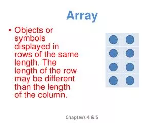
Tissue Array • Re-locating tissue from conventional paraffin or frozen tissue blocks so that multiple tissues from multiple patients can be seen on the same slide. • Originally described by in 1987 by Wan, Fortuna and Furmanski • The spots contain small tissue sections from unique tissues or tumors.
Tissue MicroArray Construction • Assembled by punching cores from specific locations in PPFE or Frozen tissue blocks and re-embedding them in a tissue array block. A section is then cut from the block and placed in a slide.
Tissue Array Construction • The current Beecher Instruments arraying device is designed to produce sample circular spots that are at least 0.6mm in diameter. • The number of spots on a single slide is variable depending on the array design. The More spots are traded off with less info per spot. • BioChain will make tissue arrays based on customer needs. Practically BioChain makes maximum 96 cores with 1.5 mm diameter.
BioChain’s Frozen Tissue Array • Benefits: • The Best quality of frozen tissue array technology and service. • Better antigen detection than Paraffin tissue section in various applications • High quality customer service
FDA Standard Frozen Tissue Array Slide 1 of both H.E. stained Image and IHC stained image by β-actin antibody. First 3 spots are adrenal from 3 different donors
Trend in changing over to Frozen Tissue Section/Array • Quote: “…I am not to happy with (PPFE tissue sections) and am contemplating changing over to frozen sections for upcoming studies…” -------A Researcher at Unilever,2007
Frozen Tissue Array Summary • Better antigen exposure-Easier to detect by IHC and ISH • Less antigen modification-Better for antigen extraction and analyses • High content- Save material cost and process time • Can be automation for Image analyses
How to sell • Understand frozen tissue array benefits • Look for pharma and biotech companies where they are developing therapeutic antibody • Check into specific labs where they are screening antibody using IHC and ISH. Histochemistry lab, Pathology lab, etc • Pre-clinical animal labs for antibody validation