Download
1 / 59
1.32k likes | 3.58k Views
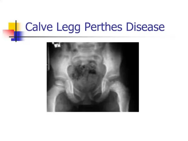
LegG -Calve- Perthes Disease. Derek Butterwick Dec 15, 2011. Perthes. Essentially idiopathic AVN of the femoral epiphysis in children. History- 1909 - 1910. Legg Calve Perthes. * 100 th Anniversary.

E N D
LegG-Calve-Perthes Disease Derek Butterwick Dec 15, 2011
Perthes • Essentially idiopathic AVN of the femoral epiphysis in children
History- 1909 - 1910 Legg Calve Perthes * 100th Anniversary
Pathogenesis*Key Event* - blood supply to head disrupted *usually > 2 infarcts before LCP develops Lateral epiphyseal vessels
Pathogenesis Osteoclastic resorption, followed by osteoblastic replacement w/ woven bone
Pathogenesis • Healing • Head soft and risk of non-spherical deformity from mechanical loads • Head must remain contained within spherical cup
Waldenstrom Stages (1922) • Radiographic description of the process • Stages • Initial • Necrosis • Fragmentation • Reossification • Healed
Waldenstrom Stages (1922) • Initial • Infarction occurs • X-rays normal – for 3 months • Necrosis • Epiphysis – dense, sclerotic • Subchondral Fracture • shows extent of necrotic epiphysis • On Lateral x-ray • a.k.a. “Crescent Sign”, “Caffery’s Sign”
Waldenstrom Stages (1922) • Fragmentation • Fissures, collapse of epiphysis • Fragmented • Extrusion of Head • Anterolateral part of epiphysis outside the lateral edge of acetabulum • > 20% extruded • risk of permanent deformity *Occurs over a 1 yr period
Waldenstrom Stages (1922) • Reossification • Creeping substitution • Occurs over 3-5 years • Healed • All woven bone, converted to lamaller bone • Assess final head deformity
End Result When Healed • Depends on how SPHERICAL the head is • Deformity following collapse • Coxa magna (large) • Coxaplana (flat) • Coxabreva (short) • Hinge abduction • Late Problems • Central Head OCDs • FAI • LLD – involved leg shorter Coxa Irregularis
Etiology - Controversial VERSUS
Etiology • Probably • Genetically susceptible child exposed to certain environment triggers • Trauma • Thrombophilia – Hot Topic • Investigated - Protein C or S deficiencies, Factor V Leiden • Nothing conclusive • Lack of modifiable factors at this point • Does this even matter to us right now?
Associations • Family History • Common - Asians, Central Europeans, Inuit • Uncommon – Blacks, Native Americans • Low birth weight, low ht. and wt. • Abnormal birth presentation • Home life – urban living, hyperactivity (i.e. ADHD), second hand smoke, older parents • Delayed bone Age
Presentation • Age – 3-8 • Symptoms • Insidious onset • Limp (painless or mild pain) x many weeks • Referred pain to distal medial thigh – 15% • Acute increase in pain • Fragmentation phase – acute collapse • Bilateral – 12% • Never simultaneous • i.e. Never same time, same stage Presents to you
Presents • Signs • Antalgic gait • Irritable hip • Pain with IR, abduction • With Head Deformity • Trendelenberg gait/sign (short abductors) • Stiff hip • tight abduction • Loss of IR • LLD – from adduction contracture
Differential Diagnosis • Anything for irritable hip • Septic Arthritis, transient synovitis, etc. • Spondyloepiphyseal dysplasia tarda • GauchersDz • AVN • Sickle cell • Steroids • Consider if • Bilateral • Symmetric • Other joints involved
Work-up • X-rays • AP/Frog leg lateral • Others • Bone Scan – pinhole • MRI • Arthrogram
Classifications • Many – I found 8! • Most important ones: • Salter-Thompson • Catterall • Herring • Stulberg Know these two!
Salter-Thompson Classification • Based on LATERAL X-RAY • Group A – crescent sign < 50% head • Group B – crescent sign > 50% head • Allows early prediction of severity
Salter-Thompson – Group A Defines area of necrosis
Salter-Thompson – Group B **Necrosis of most of the head
Catteral Classification • Based on the extent of necrotic epiphysis involved • Use during FRAGMENTATION phase • Use AP and LATERAL views • I – anterior only • II – anterior with sequestrum • III – small part of epiphysis NOT involved • IV – entire epiphysis involved • Problem – POOR INTEROBERVER RELIABILITY • Rarely used
Herring (lateral pillar) Classification • Use: AP X-RAY,Fragmentation Phase • Based on Height of Lateral Pillar • Lateral 1/3 of head • A – Normal Ht • B - > 50% of ht maintained • B/C boarder – exactly 50% of ht • C - < 50% of ht maintained • Great prognostic value • But classification made during fragmentation phase • Ideally – hip is treated before fragmentation
Herring Classification A B C
Stulberg Classification • Assess spherocity and congruity of head on healed • Ideally – all hip I or II • - *Best predictors of Long Term Prognosis (i.e. OA risk)*
Pinhole Scintigraphy • Conway Classification • Used as an early tool for diagnosis and prognostic value • Advantage – detects LCP before fragmentation stage • A Pathway – re-canalization of the lateral epiphyseal vessels • Lateral column formation – i.e. Early and rapid revascularization • Good outcome • B Pathway – neovascularization of the epiphysis • Activity central at the physi, extending laterally • Poor Outcome
Pinhole Initial Stage - Totally avascular Lateral Column formation Poor lateral column
Poor Prognostic Factors • AGE • < 6 years – GOOD PROGNOSIS – 60% Stulberg II • > 8 years – POOR PROGNOSIS – 40% Stulberg II • Extent of head involvement • Herring classification – B/C or C • Subchondral fracture - > 50% • Premature physeal closure • Poor ROM • 2 ≥ “head at risk signs”
Head at Risk Signs • Lateral Subluxation • Calcification lateral to the epiphysis • Diffuse metaphyseal reaction • Horizontal growth plate • Gage sign – V shaped bone cyst in lateral femoral head
Treatment Rules • Any Herring C Hips • All do poorly - no matter what age or treatment • 13% chance of good result (Stulberg I/II) • Under 6 • All do well - except lateral pillar C • Observe + Symptomatic treatment • Those > age 8 • Herring A – do well • B or B/C – benefit from surgical treatment • i.e. B – surgery (73% Stulberg I/II hip), no surgery (44% Stulberg I/II) • Age 6-8 • With B or B/C boarders – controversial • Treat if poor prognostic factors
Who can you observe? • Any Child < 6 yrs old • All Lateral Pillar A Hips • Treatment • Symptomatic treatment • Physiotherapy – maintain ROM (abduction of 20 degrees), nourishes cartilage • Consider avoiding impact activities during fragmentation phase • Think of as self limiting disorder • *All others consider treatment*
Treatment • TREATMENT = CONTAINMENT • Epiphysis is completely within the acetabulum, which shapes head while epiphysis is healing • Extrusion = lack of containment • Containment allows for congruence • The earlier the head is contained in disease, the better the results • For use before the reossification stage • Short Term Goals • CONTAINMENT • MAINTAIN ROM • Long Term Goals • Obtain spherical healed head – avoid OA
Candidate for Containment • Any child > 8 years old with • Lateral pillar B, B/C, C • Catterall III, IV • Salter-Thompson B
Casts For Containment Petrie Cast • Abduction, IR casting • Sometimes require • Adductor tenotomy to increase abduction • Weight bearing – allow flexion/extension of hips • Stimulates remodelling • D/C cast – when lateral pillar re-ossifies • In general – rarely used
Braces for Containment Results Atlanta Abduction Orthoses • Poor Results • No better than observation + ROM exercises • Hard to control rotation of the leg • Abandoned!!!
Why non-operative? • If surgery offers better results for B & B/C boarder hip, then why cast?
Surgical Containment Femoral Varus Osteotomy InnominateOsteotomy
Surgical Containment • Equal results with either osteotomy • Alters natural history of disease • May bypass or shortening fragmentation phase • Extent of extrusion decreased
Femoral Varus Osteotomy • Better in early clinical stage • i.e. Necrosis or early fragmentation • Intra-operative arthrography – find best containment position • Subtrochanteric Osteotomy • Average chance in neck shaft angle = 30 degrees • Must have enough abduction remaining after osteotomy
Femoral Varus Osteotomy Advantages Disadvantages • Technically easier • Decreases pressure on head • Increases LLD • Average shortening 1 cm • Permanent CoxaVara • Abductor insufficiency (trendelenberg) • Hardware removal
Salter Osteotomy • Provides anterolateral coverage • Acetabulum displaced medially – decreased pressure on head • Acetabulum displaced distally – increases leg length
Salter Osteotomy Advantages Disadvantages • Does not shorten neck • Restores leg length • Improves trendelenberg gait • Easier hardware removal • Technically more demanding • Requires more experience
Hinge Abduction • Usually seen in late ossification or healed stages • Abnormal Hip Mechanics • Extruded epiphysis impinges and hinges on lateral acetabulum • Painful • Progressive – collapse of lateral pillar, flattening of the head, and subluxation • Abandon containment goal Entire Epiphysis not containable New Treatment Goal - CONGRUENCY
Hinge Abduction Arthrogram Abduction View Adduction View Congruent “Position of Best Fit” Medial dye pooling Incongruent Hip Hinge Abduction
Proximal Femoral Valgus Osteotomy • Vaglusosteotomy – place head in position of best fit, where hip is most congruent • Usually posteromedial head • Advantages • Pain relief ...lateral epiphysis not impinging • Congruent joint ...less point loading, less OA • Improves LLD ...from collapse of femoral head • Re-tensions Abductors ...reduces tredelenberg • Allows acetabular remodelling laterally • Disadvantages • Abduction contracture...if abductors shortened previously • No Valgus > 160 degrees
Case Example get lateral acetabular growth (coverage) get remodelling of epiphysis bump