Download
1 / 19
360 likes | 2.2k Views
Phonons and Inelastic neutron and X-ray Scattering Paolo Ghigna, Dipartimento di Chimica Fisica “M. Rolla”, Università di Pavia Summary

E N D
Phonons and Inelastic neutron and X-ray Scattering Paolo Ghigna, Dipartimento di Chimica Fisica “M. Rolla”, Università di Pavia
Summary • The study of atomic dynamics in condensed matter at momentum transfers, Q, and energies, E, characteristic of collective motions is, traditionally, the domain of neutron spectroscopies. • The experimental observable is the dynamic structure factor S(Q,E), which is the space and time Fourier transform of the density-density correlation function. • Neutrons as probing particle are particularly suitable, since • the neutron-nucleus scattering cross-section is sufficiently weak to allow for a large penetration depth, • the energy of neutrons with wavelengths of the order of inter-particle distances is about 100 meV, and therefore comparable to the energies of collective excitations associated to density fluctuations such as phonons, and • the momentum of the neutron allows to probe the whole dispersion scheme out to several Å-1, in contrast to inelastic light scattering techniques such as Brillouin and Raman scattering which can only determine acoustic and optic modes, respectively, at very small momentum transfers.
What is a phonon? • In physics, a phonon is a quantizedmode of vibration occurring in a rigid crystal lattice, such as the atomic lattice of a solid. Phonon can also be used to describe an exitation of such a mode. The study of phonons is an important part of solid state physics, because phonons play an important role in many of the physical properties of solids, such as the thermal conductivity and the electrical conductivity. In particular, the properties of long-wavelength phonons gives rise to sound in solids -- hence the name phonon. In insulating solids, phonons are also the primary mechanism by which heat conduction takes place. • Phonons are a quantum mechanical version of a special type of vibrational motion, known as normal modes in classical mechanics, in which each part of a lattice oscillates with the same frequency. These normal modes are important because, according to a well-known result in classical mechanics, any arbitrary vibrational motion of a lattice can be considered as a superposition of normal modes with various frequencies; in this sense, the normal modes are the elementary vibrations of the lattice. Although normal modes are wave-like phenomena in classical mechanics, they acquire certain particle-like properties when the lattice is analysed using quantum mechanics (see wave-particle duality.) They are then known as phonons. Phonons are bosons possessing zero spin.
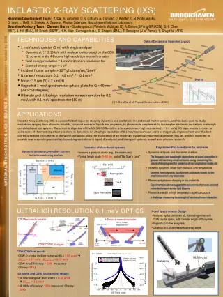
X-rays as a probe of phonons • While it has been pointed out that X-rays can in principle as well be utilised to determine the S(Q,E), it was stressed that this would represent a formidable experimental challenge, mainly due to the fact that an X-ray instrument would have to provide an extremely high energy resolution. This is understood considering that photons with a wavelength of l=0.1 nm have an energy of about 12 keV. Therefore, the study of phonon excitations in condensed matter, which are in the meV region, requires a relative energy resolution of at least DE/E=10-7. • On the other hand, there are situations where the use of photons has important advantages over neutrons. A specific case is based on the general consideration that it is not possible to study acoustic excitations propagating with a speed of sound vs using a probe particle with a speed v smaller than vs. • This limitation is not particularly relevant in neutron spectroscopy studies of crystalline samples. Here, the translation invariance allows to study the acoustic excitations in high order Brillouin zones, thus overcoming the above mentioned kinematic limit on phonon branches with steep dispersions. • On the contrary, the situation is very different for topologically disordered systems such as liquid, glasses and gases. In these systems, in fact, the absence of periodicity imposes that the acoustic excitations must be measured at small momentum transfers. Thermal neutrons have a velocity in the range of 1000 m/s, and only in disordered materials with a speed of sound smaller than this value (mainly fluids of heavy atoms and low density gases) the acoustic dynamics can be effectively investigated. • Another advantage of the inelastic X-ray technique arises from the fact that very small beam sizes of the order of a few tens of micrometers can be presently obtained at third generation synchrotron sources. This allows to study systems available only in small quantities down to a few 10-6 mm3 and/or their investigation in extreme thermodynamic conditions, such as very high pressure. These differences with respect to inelastic neutron scattering motivated the development of the very high resolution inelastic x-ray scattering (IXS) technique, and following the pioneering experiments in 1986, the IXS technique rapidly evolved. To date there are four instruments operational at the ESRF (2), APS (1) and Spring-8 (1), and several more under construction.
What is a phonon? • Due to the connections between atoms, the displacement of one or more atoms from their equilibrium positions will give rise to a set of vibration waves propagating through the lattice. One such wave is shown in the figure below. The amplitude of the wave is given by the displacements of the atoms from their equilibrium positions. The wavelength λ is marked. • It should be noted that there is a minimum possible wavelength, given by the equilibrium separation a between atoms. As we shall see in the following sections, any wavelength shorter than this can be mapped onto a wavelength longer than a. • Not every possible lattice vibration has a well-defined wavelength and frequency. However, the normal modes (which, as we mentioned in the introduction, are the elementary building-blocks of lattice vibrations) do possess well-defined wavelengths and frequencies. We will now examine these normal modes in some detail.
Inelastic Scattering • We will consider inelastic scattering where there is a change in the energy of the scattered beam with respect to the incident beam due to interactions of the incident wave with the sample. • This has proved to be a fruitful area of investigation, particularly with neutrons where the energy of thermalised neutrons is comparable to that of phonons. This was recently recognised in the award of the 1995 Nobel Prize in Physics.
Optical, Acoustic, Transverse, Longitudinal Phonon • In real solids, there are two types of phonons: "acoustic" phonons and "optical" phonons. "Acoustic phonons", which are the phonons described above, have frequencies that become small at the long wavelengths, and correspond to sound waves in the lattice. Longitudinal and transverse acoustic phonons are often abbreviated as LA and TA phonons, respectively. • "Optical phonons," which arise in crystals that have more than one atom in the unit cell, always have some minimum frequency of vibration, even when their wavelength is large. They are called "optical" because in ionic crystals (like sodium chloride) they are excited very easily by light (in fact, infrared radiation). This is because they correspond to a mode of vibration where positive and negative ions at adjacent lattice sites swing against each other, creating a time-varying electrical dipole moment. Optical phonons that interact in this way with light are called infrared active. Optical phonons which are Raman active can also interact indirectly with light, through Raman scattering. Optical phonons are often abbreviated as LO and TO phonons, for the longitudinal and transverse varieties respectively.
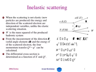
High Energy X-ray Inelastic Scattering • Ever since the epoch-making DuMond experiments on beryllium, which provided the first evidence for the validity of Fermi-Dirac as opposed to Maxwell-Boltzmann electron momentum distributions, inelastic x-ray scattering has been established as a probe of the ground state properties of electrons in solids. • Inelastic scattering refers to a number of interactions between x-rays and atoms in which the energy of the scattered photon is less than that of the incident one. The term high energy indicates the relative magnitude of the incident photon energy in comparison to the electron binding energy. • Amongst the effects are the Compton effect, plasmon scattering (or collective excitation), x-ray Raman, x-ray resonant Raman and phonon scattering. In a typical scattering experiment an incident beam of photons of energy hn1 and wavevector k1is scattered by the sample into a beam of photons of average energy hn2 and wavevector k2. The spectral distribution then provides information about the electronic structure of the sample. • One advantage of this type of experiment is that the scattering probe is a quasi-particle of energy hn = h(n1-n2 ) and momentum (h/2p)k = (h/2p)(k1-k2) and the functional dependence between kand wis multi-valued and can be “tuned” appropriately to study a desired interaction. Large momentum transfers correspond to Compton scattering providing information on the ground state momentum of the individual electrons.
High Energy X-ray Inelastic Scattering • Intermediate and low momentum transfer are governed by the differential scattering cross section • where e(w,k) is the dynamic dielectric function and S(w,k), the dynamic structure factor provides information about the correlation of the valence electrons in space and time. For the core electrons x-ray Raman spectra provide information about the density of states near the Fermi energy and many body effects while x-ray resonant Raman and phonon inelastic scattering can provide details of the spin dependent momentum distribution
X-ray Inelastic Scattering DuMond (in 1929!) developed from first principles, a relation between the electron momentum distribution I(p) of an isotropic ensemble of electrons and the spectrum of a monochromatic x-ray beam inelastically scattered by them. This relation which leads to: DuMond’s experimental spectra of Be were clearly most similar to (i) which was the first direct evidence for the validity of the Fermi-Dirac electron momentum distribution.
Inelastic Neutron Scattering • This is the principle technique for determining phonon dispersion curves and is of major importance in the study of phonons generally. • Thermal neutrons (i.e. those in the meV energy range) have both the right energy and wavevector to interact with phonons. This is in marked contrast to other probes such as infrared radiation (where the energy is similar but (h/2p)k is only a very small fraction of the Brillouin zone boundary, and hence only phonons with k0 can be measured) or x-rays ( where (h/2p)k can be of the correct order but the energy is many orders of magnitude larger than that of phonons). • Note that a lattice vibration of angular vibration wconnotes the movement of phonons, each with energy hnand crystal momentum (h/2p)k . Use of the term crystal momentum does not mean that ordinary momentum is transferred through the crystal at the corresponding rate, but “crystal momentum” is a conserved quantity in interactions involving the creation or annihilation of crystal phonons.
Inelastic Neutron Scattering • A neutron of velocity v has a wavevector and kinetic energy • Suppose that in either absorbing or creating a phonon, the energy and wavevector of the neutron are changed to E’ and Kn’ respectively. Then the angular frequency w and wavevector k of the phonon involved will be related by the conservation equations • The vector G is either zero or a reciprocal lattice vector and can be included since a phonon of wavevector k is identical to a phonon of any (k+G). The positive signs in are for the creation of a phonon (Stokes process) and the negative signs for the annihilation (anti-Stokes) of a phonon.
Triple-axis spectrometer • The beam of neutrons is monochromated to the desired wavelength via Bragg reflection of a crystal monochromator. After the beam is scattered by the sample an analyser crystal is adjusted so that it Bragg reflects a defined energy of the scattered neutrons towards the detector. • In the triple-axis spectrometer the incident energy can be kept constant by keeping qMconstant. The scattered energy is adjusted by changing qA, but the consequent change in ½k’½ can be compensated by simultaneously altering the scattering angle fto keep ½K½=½k’-k½ constant; also the angle yof the sample can be varied so that K maintains its orientation with respect to the crystal axes. Thus by simultaneously changing the three angles qA, fand y, it is possible to investigate the variation of neutron count rate with the energy E’ of the scattered neutrons at fixed values of the incident neutron energy E and scattering vector K.
Phonon dispersion curves • The technique can thus be applied to measuring the phonon frequencies as a function of wavevector throughout the Brillouin zone of the material. The figures display the results for two f.c.c metals and for f.c.c silicon which has two atoms in the basis and thus displays both acoustic and optic branches in the Brillouin zone
Comparison IXS versus INS • Similarities and differences between the IXS and INS cross section are summarised below: • Q - E limitation for INS • Small beam size for IXS • X-rays couple to the electrons of the system with a cross-section proportional to the square of the classical electron radius, ro=2.82x10⁻¹³ cm, i.e. with a strength comparable to the neutron-nucleus scattering cross-section b. • The IXS cross section is proportional to fj(Q)2. In the limit Q0, the form factor is equal to the number of electrons in the scattering atom, Z; for increasing values of Q, the form factor decays with decay constants of the order of the inverse of the atomic wavefunction dimensions of the electrons in the atom. • The total absorption cross-section of X-rays above 10 keV energy is limited in almost all cases (Z>4) by the photoelectric absorption process, and not by the Thomson scattering process. The photoelectric absorption, whose cross-section is roughly proportional to Z4, determines therefore the actual sample size along the scattering path. Consequently the Thomson scattering channel is not very efficient for system with high Z in spite of the Z2 dependence of its cross-section. • As a consequence, multiple scattering processes can in general be neglected • The magnetic cross section is negligible for IXS, whereas it is comparable to the nuclear cross section for neutrons. • The IXS cross section is highly coherent • The shape of the IXS instrumental energy resolution is not Gaussian as it is for a neutron triple-axis spectrometer, but Lorentzian.
General optical lay-out • The optical lay-out is based on the triple-axis principle, composed of the very high energy resolution monochromator (first axis), the sample goniometry (second axis) and the crystal analyser (third axis). The figure below shows a schematic view of the optics and the distances involved. Due to the backscattering geometry the beamline is fairly long in order to acquire a sufficient beam offset between the incident photon beam from the X-ray source and the focused very high-energy resolution beam at the sample position
Backscattering monochromator • The main monochromator consists of a flat perfect single crystal, operating at a Bragg angle of 89.98º and utilising the silicon (n,n,n,) reflection orders. This close-to-exact-backscattering configuration insures that the spectral angular acceptance, the so-called Darwin width, is larger than the x-ray beam divergence, and therefore that all the photons within the desired energy bandwidth are transmitted. The crystal is temperature controlled in the mK region by a high precision platinum 100W (Pt100) thermometer bridge in closed-loop operation with a PID controlled heater unit. The complete electronic unit was purchased from Automatic Systems Laboratory (Milton Keynes, England). • The energy scans are performed by varying the temperature of the monochromator and keeping the temperature of the analyzer fixed.
Very high energy resolution spherical crystal analyzer • Although the required energy resolution is the same for the monochromator and the analyser, there is a big difference concerning their angular acceptance. The spatial angular acceptance of the monochromator should be compatible with the divergence of the synchrotron beam, whereas the analyser must have a much larger angular acceptance, which is only restricted by the required Q resolution. An angular acceptance up to 4x10mrad2 is an adequate compromise of Q-resolution and signal maximisation. These constraints necessitate focusing optics. Since it is not possible to elastically bend a crystal without introducing important elastic deformations, which in turn deteriorate the intrinsic energy resolution, one has to position small, unperturbed crystals onto a spherical substrate with a radius, fulfilling the Rowland condition. This polygonal approximation to the spherical shape yields the intrinsic energy resolution, provided the individual crystals are unperturbed and the geometrical contribution of the cube size to the energy resolution is negligible. The solution realised at the ESRF consists of gluing 12000 small crystals of 0.6x0.6x3 mm3 size onto a spherical silicon substrate. This procedure, yield very good results, and provided the record energy resolution of 0.9 meV, utilising the silicon (13,13,13) reflection order at 25704 eV.