Download
1 / 21
220 likes | 978 Views
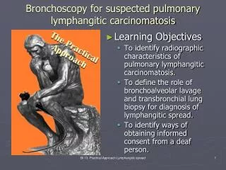
Bronchoscopy for suspected pulmonary lymphangitic carcinomatosis. Learning Objectives To identify radiographic characteristics of pulmonary lymphangitic carcinomatosis. To define the role of bronchoalveolar lavage and transbronchial lung biopsy for diagnosis of lymphangitic spread.

E N D
Bronchoscopy for suspected pulmonary lymphangitic carcinomatosis • Learning Objectives • To identify radiographic characteristics of pulmonary lymphangitic carcinomatosis. • To define the role of bronchoalveolar lavage and transbronchial lung biopsy for diagnosis of lymphangitic spread. • To identify ways of obtaining informed consent from a deaf person. BI 13. Practical Approach Lymphangitic spread
G.G. is a 72 year old man with stage IV adenocarcinoma of the lung admitted for progressive dyspnea. He has undergone multiple chemotherapy regimens. Four months before admission he was started on tyrosine kinase inhibitors. He has increasing shortness of breath, fatigue, dry cough, and weight loss for several weeks. The patient has COPD with FEV1 35% predicted and is deaf. He lives with his 33 year old son. Karnoksky status is 50. Chest radiograph shows diffuse bilateral interstitial infiltrates and an ill-defined opacity at the right lung base. Computed tomography scan reveals intralobular septal thickening and consolidation in the right middle lobe which was the site of the primary tumor. The oncology team has formulated a differential diagnosis that includes lymphangitic carcinomatosis, pulmonary infection, and drug-related pneumonitis. Pulmonary consultation is requested for bronchoscopy Case description BI 13. Practical Approach Lymphangitic spread
Initial Evaluation Procedural Strategies Techniques and Results Long term Management The Practical Approach • Examination and, functional status • Significant comorbidities • Support system • Patient preferences and expectations • Indications, contraindications, and results • Team experience • Risk-benefits analysis and therapeutic alternatives • Informed Consent • Anesthesia and peri-operative care • Techniques and instrumentation • Anatomic dangers and other risks • Results and procedure-related complications • Outcome assessment • Follow-up tests and procedures • Referrals • Quality improvement BI 13. Practical Approach Lymphangitic spread
Initial Evaluation • Physical Exam • T 37.6 BP 112/74 P 92 R 22 SaO2 91% RA • General: NAD but ill-appearing, A&Ox4, cachectic • HEENT: PERRLA, sclera anicteric, no neck LAD • Chest: diffuse bilateral crackles with decreased BS at right lung base, no wheezing • Heart: RRNR S1 S2 no murmur • Abd: soft, NT, ND, normoactive BS • Ext: +digital clubbing, no edema or cyanosis • Labs • Chem panel: Na+ 136 BUN 33 Crt 1.7 Glu 124 • CBC: WBC 12.3 Neutrophil 78% no bands Hgb 13.3 Plt 163 • Blood cx NGTD, U/A neg, sputum cx pending (Gram stain neg) BI 13. Practical Approach Lymphangitic spread
Initial Evaluation • Left: chest radiograph shows diffuse bilateral interstitial infiltrates and an ill-defined opacity at the right lung base. Right: computed tomography scan reveals intralobular septal thickening and consolidation in the right middle lobe which was the site of the primary tumor. BI 13. Practical Approach Lymphangitic spread
Initial Evaluation • Functional status assessment • Karnofsky status score 50 • Significant co-morbidities • Advanced COPD, HTN, chronic renal insufficiency, poor functional capacity • Support system • Lives with wife, has 3 children who are supportive • Preferences and expectations • Realistic and understands severity of disease BI 13. Practical Approach Lymphangitic spread
Karnofsky Performance Status Scale Definitions Rating (%) Criteria • Able to carry on normal activity and to work; no special care needed • 100: normal; no complaints; no evidence of disease • 90: able to carry on normal activity; minor signs or symptoms of disease • 80: normal activity with effort; some signs or symptoms of disease • Unable to work; able to live at home and care for most personal needs; varying amount of assistance needed • 70: cares for self; unable to carry on normal activity or to do active work • 60: requires occasional assistance, but is able to care for most of his personal needs • 50: requires considerable assistance and frequent medical care • Unable to care for self; requires equivalent of institutional or hospital care; disease may be progressing rapidly • 40: disabled; requires special care and assistance • 30: severely disabled; hospital admission is indicated although death not imminent • 20: very sick; hospital admission necessary; active supportive treatment necessary • 10: moribund; fatal processes progressing rapidly • 0: dead BI 13. Practical Approach Lymphangitic spread
Procedural Strategies • Indications for bronchoscopy • Diagnosis of lymphangitic carcinomatosis • Evaluation of presence of infection • Bacterial vs. fungal vs. viral • Contraindications for bronchoscopy • No history of recent MI or arrhythmia • History of advanced COPD • 5% of COPD patients with bronchoscopy-related complication compared to 0.6% in patients with normal lung function • Especially at risk: • FEV1/FVC <50% or • FEV1 <1L and FEV1/FVC <69% • Consider pre-procedure spirometry in severe COPD (increased concern if FEV1 <40%) • Use sedation and O2 carefully in patients with elevated CO2 (concern for retention) BI 13. Practical Approach Lymphangitic spread
Procedural Strategies • Operator and team experience • Risks of bronchoscopy • Mortality rate: 0.01-0.04% • Complication rate: 0.12-0.30% • Oversedation • May cause desaturations or CO2 retention • Bronchospasm • Premedicate with bronchodilator to reduce decrease in FEV1 • Data in asthmatic patients • Laryngospasm • Hypoxemia • Maintain SaO2 > 90% in perioperative setting • Bleeding • UCI policy is to check preoperative CBC and coagulation panel • BTS recommends routine preop coags before bronchoscopy BI 13. Practical Approach Lymphangitic spread
Procedural Strategies • Informed consent for the hearing-impaired • When deaf patients sign consent forms, they often do so without understanding them • Many believe forms are malpractice waivers • Involvement of an interpreter is indispensable • Allows physician to • Obtain thorough history and proper examination • Ensure that patient understands risks and benefits of procedure • Establish effective communication and environment of caring and trust • Communicate through writing when possible as opposed to lip-reading BI 13. Practical Approach Lymphangitic spread
Procedural Techniques and Results • Anesthesia: moderate sedation • Midazolam • Well tolerated • Less than 10% exhibit prolonged effect from impaired metabolism • Initial dose of 2 mg followed after 2 minutes by increments of 1 mg/min if needed • Propofol: monitor for hypotension • Flumazenil: benzodiazepine antagonist • Short elimination time allows re-sedation • Usual initial dosage: 250-500 micrograms • Perioperative care • Continuous oxygen with monitoring • Preoperative bronchodilator • Viscous and liquid Lidocaine • Procedure-related complications *reference BI 13. Practical Approach Lymphangitic spread
Long-term Management Plan • BAL was performed and revealed malignant cells consistent with adenocarcinoma • Diagnosis of lymphangitic carcinomatosis was discussed with patient and given poor prognosis a palliative care consult was considered • Patient was discharged home with hospice • Patient and family expressed satisfaction with care and management by all subspecialty and ancillary teams BI 13. Practical Approach Lymphangitic spread
Q 1: What are the specific CT characteristics of pulmonary lymphangitic carcinomatosis and how do they differ from those of tyrosine kinase inhibitor-induced interstitial pneumonitis? • HRCT findings in lymphangitic carcinomatosis • Irregular, nodular, and/or smooth interlobular septal thickening • Thickening of fissures as result of involvement of lymphatics concentrated in subpleural interstitium • Preservation of normal parenchymal architecture at level of second pulmonary lobule • Peribronchovascular thickening • Centrilobular peribronchovascular thickening predominating over interlobular septal thickening in a minority of patients • Polygonal arcades or polygons with prominence of centrilobular bronchovascular bundle in association with interlobular septal thickening (50%) • Mediastinal and/or hilar lymphadenopathy (30-50%) • Pleural effusions (30-50%) • Findings can be unilateral or bilateral and focal or diffuse BI 13. Practical Approach Lymphangitic spread
Q 1: What are the specific CT characteristics of pulmonary lymphangitic carcinomatosis and how do they differ from those of tyrosine kinase inhibitor-induced interstitial pneumonitis? • HRCT findings in tyrosine kinase-induced interstitial pneumonitis • Diffuse interstitial markings and increased radiodensities • Ground glass opacities • Multiple centrilobular nodules • Focal air trapping • Pleural effusion • Extensive fibrosis and honeycombing with traction bronchiectasis in chronic and advanced disease BI 13. Practical Approach Lymphangitic spread
Lymphangitic carcinomatosisSmooth and nodular interlobular septal thickening (black arrow) BI 13. Practical Approach Lymphangitic spread
Lymphangitic carcinomatosisProminent axial interstitium with thickened bronchovascular bundles (solid arrow). Also thickening of fissure secondary to involvement of subpleural lymphatics (open arrow). BI 13. Practical Approach Lymphangitic spread
Tyrosine kinase-induced interstitial pneumonitis BI 13. Practical Approach Lymphangitic spread
Q 2: What is the expected yield of bronchoalveolar lavage for diagnosing lymphangitic carcinomatosis, and how does this yield compare with that of transbronchial lung biopsy? • Transbronchial lung biopsy • Goal is to replace more invasive open lung or transthoracic needle biopsy • 19 patients with diffuse interstitial disease underwent flexible bronchoscopy with transbronchial biopsy • Lymphangitic carcinomatosis was established in 6 (32%) of patients • One patient developed 30% pneumothorax which was treated with chest tube evacuation • The diffuse bronchial and peribronchial lymphatic involvement demonstrated suggests that TBLB should be the procedure of choice in diagnosis of lymphangitic carcinomatosis • Aranda C et al. Transbronchial lung biopsy in the diagnosis of lymphangitic carcinomatosis. Cancer 1978;42:1995-8. BI 13. Practical Approach Lymphangitic spread
Q 2: What is the expected yield of bronchoalveolar lavage for diagnosing lymphangitic carcinomatosis, and how does this yield compare with that of transbronchial lung biopsy? • Bronchoalveolar lavage • Retrospective analysis, 12 patients with known neoplastic disease and diffuse pulmonary infiltrates consistent with lymphangitic carcinomatosis • BAL correctly identified 5/5 (100%) • No complications • Bronchial washing 4/7 (57%) • Bronchial brushing 2/5 (40%) • Transbronchial lung biopsy 4/9 (44%) • One patient with significant pulmonary hemorrhage • Conclusion: BAL should be performed to confirm diagnosis before proceeding to biopsy, especially when risks of pneumothorax and hemorrhage are excessive • The value of bronchial washings and bronchoalveolar lavage in the diagnosis of lymphangitic carcinomatosis. Chest 1998;94:1028-30 ref. BI 13. Practical Approach Lymphangitic spread
Q 3: Should you perform transbronchial lung biopsy in this case? If so, why? If not, why not? • History of advanced COPD with increased concern for pneumothorax • History of chronic renal insufficiency with concern for platelet dysfunction and increased risk of bleeding • Evidence showing BAL as the initial test of choice in diagnosis of lymphangitic carcinomatosis • Consider TBLB if BAL is non-diagnostic and patient wishes to pursue further diagnosis and treatment BI 13. Practical Approach Lymphangitic spread
Prepared with the assistance of Paul Huang M.D. www.bronchoscopy.org BI 13. Practical Approach Lymphangitic spread