Download
1 / 1
30 likes | 257 Views
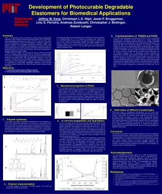
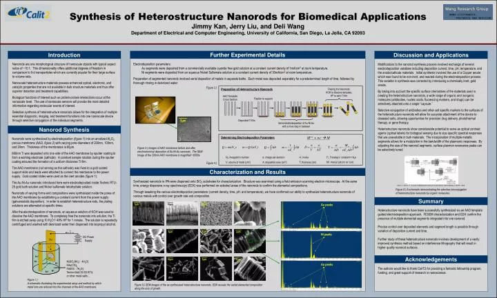
Freeing the Nanorods: KOH to dissolve template, HF to etch Ti film. AAO Template Cross Section. Fixation to support. 5 μ m. Deposited Ti film. Serial electrodeposition of Au-Ni-Au with a rinse step in between. Synthesis of Heterostructure Nanorods for Biomedical Applications

E N D
Freeing the Nanorods: KOH to dissolve template, HF to etch Ti film AAO Template Cross Section Fixation to support 5μm Deposited Ti film Serial electrodeposition of Au-Ni-Au with a rinse step in between Synthesis of Heterostructure Nanorods for Biomedical Applications Jimmy Kan, Jerry Liu, and Deli Wang Department of Electrical and Computer Engineering, University of California, San Diego, La Jolla, CA 92093 Further Experimental Details Introduction Discussion and Applications • Electrodeposition parameters: • Au segments were deposited from a commercially available cyanide free gold solution at a constant current density of 1mA/cm2 at room temperature. • Ni segments were deposited from an aqueous Nickel Sulfamate solution at a constant current density of 30mA/cm2 at room temperature. • Preparation of segmented nanorods involved serial deposition of metals in separate baths. Each metal was deposited separately for a predetermined length of time, followed by thorough rinsing in deionized water. Nanorods are one morphological structure of nanoscale objects with typical aspect ratios of ~10:1. This dimensionality offers additional degrees of freedom in comparison to 0-d nanoparticles which are currently popular for their large surface to volume ratio. Nanoscale heterostructure materials possess enhanced optical, electronic, and catalytic properties that are not available in bulk structure materials and thus offer superior detection and treatment capabilities. Biological functions of interest such as protein-protein interactions occur at the nanoscale level. The use of nanoscale sensors will provide the most detailed information regarding molecular events of interest. Selective synthesis of heterostructure nanorods allows for the integration of multiple essential diagnostic, imaging, and treatment functions into one nanoscale device through selective conjugation of the individual segments. Modifications to the nanorod synthesis process involved exchange of several electrodeposition variables including deposition current, time, pH, temperature, and the anode/cathode materials. Initial synthesis involved the use of a Copper anode which was found to be non-inert, and reacted during the electrodeposition process. This variable in synthesis was corrected by introducing a chemically inert, gold anode. By taking into account the specific surface chemistries of the materials used in creating the heterostructure nanorods, a wide range of organic and inorganic molecules (antibodies, nucleic acids, fluorescing markers, and drugs) can be selectively attached onto a single “capsule.” Selective conjugation of antibodies and other cell specific markers to the surfaces of the heterostructure nanorods will allow for accurate attachment of the device to diseased cells, allowing opportunities for precision drug delivery, photothermal therapy, or gene therapy. Heterostructure nanorods show considerable potential to serve as optical contrast agents (optical labels) for biological sensing due to size specific spectral responses that are unavailable in bulk materials. The incorporation of multiple metallic segments allows for a modulation in the bandwidth of the plasmonic responses. By adjusting the size of the nanorod segments, surface plasmon resonance peaks can be selectively tuned. Figure 3.) Preparation of Heterostructure Nanorods Nanorod Synthesis Nanorods were synthesized by electrodeposition (figure 1) into an anodized Al2O3 porous membrane (AAO, figure 2) with varying pore diameters of 200nm, 100nm, and 20nm. Thickness of the membranes is 60μm. 600nm Ti was deposited onto one side of the AAO membrane by sputter coating to form a working electrode (cathode). A constant sample rotation during the sputter coating ensured the formation of a uniform thickness Ti film. The AAO membrane (not serving as the cathode) was fixed to a gold coated support slide and leads were attached to connect the membrane to the power supply. Gold coated slides were used as the inert anodes (figure 1). The Au-Ni-Au nanorods introduced here were electrodeposited inside Technic RTU-25 gold bath solution and Nickel sulfamate tetrahydrate solution. Nanorods of varying forms and compositions were synthesized inside the pores of the AAO membrane by establishing a constant current from the power supply (galvanostatic deposition). In order to establish heterostructure rods, the plating solutions are alternated at specific times. After the electrodeposition of nanorods, an aqueous solution of KOH was used to dissolve the AAO membrane. To completely free the nanorods into solution, the Ti film is etched away using 10 H2O:1 49% HF for 1 minute. The solution is repeatedly centrifuged and washed with deionized water then dispersed into isopropyl alcohol. M+n + ne- M Determining Electrodeposition Parameters Figure 2.) Images of AAO membrane before and after electrochemical deposition of Au-Ni-Au nanorods. The SEM Image of the 200nm AAO membrane is magnified ~6000x. Figure 4.) Characterization and Results Synthesized nanorods in IPA were dispersed onto SiO2 substrates for characterization. Structure was examined using a field emission scanning electron microscope. At the same time, energy dispersive x-ray spectroscopy (EDX) was performed on selected areas of the nanorods to confirm the elemental compositions. Through tweaking the various electrodeposition parameters (current density, time, pH, and temperature), we have confirmed our ability to synthesize heterostructure nanorods of various metals with control over growth rate and composition. Salem, A., Searson, P., & Leong, K. Multifunctional nanorods for gene delivery. Nature Materials 2, 668-671 (2003). Figure 6.) A schematic demonstrating the selective bioconjugation of heterostructure nanorods by organic molecules. Summary 5µm 5µm 10µm 50µm Au peaks Heterostructure nanorods have been successfully synthesized via an AAO template guided electrodeposition approach. FESEM characterization and EDX confirm the presence of multiple elemental segments integrated into one nanorod. Precise control over deposited elements and segment length is possible through variation of deposition current and time. Further study of these heterostructure nanorods involves development of a vastly improved synthesis method based on interference lithography that will result in higher quality nanorod surfaces. e - 10µm 5µm 5µm 2µm Ni peaks DC Power Supply Au e - Au+ Acknowledgements 10µm 5µm 2µm 2µm Ni(SO3NH2) · 4H2O] KAu(CN)2 FeSO4 · 7H2O] Techni-Gold 25 ES RTU, or other metal salts… Ni+ Ni+ Au peaks Ni+ Au+ Figure 1.) A schematic illustrating the experimental setup and method by which metal ions are reduced into the channels of the AAO membrane. Figure 5.) SEM images of the as synthesized heterostructure nanorods. EDX reveals the varied elemental composition along the axis of growth.