Download
1 / 32
590 likes | 1.63k Views
Nuclear Magnetic Resonance (NMR) Spectroscopy Structure Determination Introduction Theory of NMR spectroscopy Chemical shift 1 H NMR—Number of Signals 1 H NMR—Position of Signals 1 H NMR—Chemical Shift Values 1 H NMR—Intensity of Signals 1 H NMR—Spin-Spin Splitting. NMR Spectrometer.

E N D
Nuclear Magnetic Resonance (NMR) Spectroscopy • Structure Determination • Introduction • Theory of NMR spectroscopy • Chemical shift • 1H NMR—Number of Signals • 1H NMR—Position of Signals • 1H NMR—Chemical Shift Values • 1H NMR—Intensity of Signals • 1H NMR—Spin-Spin Splitting
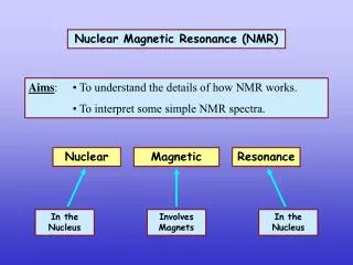
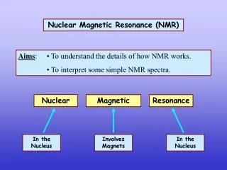
Introduction to NMR Spectroscopy • Nuclear magnetic resonance spectroscopy is a powerful analytical technique used to characterize organic molecules by identifying carbon-hydrogen frameworks within molecules. • The source of energy in NMR is radio waves which have long wavelengths, and thus low energy and frequency. • When low-energy radio waves interact with a molecule, they can change the nuclear spins of some elements, including 1H and 13C.
Only magnetic nuclei that contain odd mass numbers (such as 1H1, 13C6, 19F9 and 31P15) or odd atomic numbers (such as 2H1 and 14N7) give rise to NMR signals. • Non magnetic nuclei that have even atomic number, and even atomic mass are NMR inactive. • (12C6, 16O8) • Two common types of NMR spectroscopy are used to characterize organic structure: • A) 1H NMR is used to determine the type and number of H atoms in a molecule. • B)13C NMR is used to determine the type of carbon atoms in the molecule.
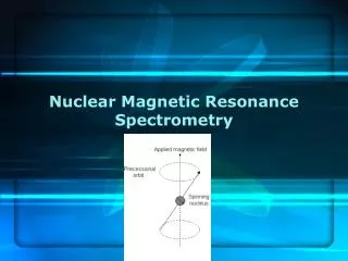
Theory of NMR Spectrum • When a charged particle such as a proton spins on its axis, it creates a magnetic field. Thus, the nucleus can be considered to be a tiny bar magnet. • Normally, these tiny bar magnets are randomly oriented in space. However, in the presence of a magnetic field B0, they are oriented with or against this applied field. More nuclei are oriented with the applied field because this arrangement is lower in energy.
In a magnetic field, there are now two energy states for a proton: a lower energy state with the nucleus aligned in the same direction as B0, and a higher energy state in which the nucleus aligned against B0. • When an external energy source (hv) that matches the energy difference (ΔE) between these two states is applied, energy is absorbed, causing the nucleus to “spin flip” from lower energy state to the higher. When the nuclei fall back to their lower energy state, the detector measures the energy released, and a spectrum is recorded.
Nuclei in different environments absorb at slightly different frequencies, so they are distinguishable by NMR. • The frequency at which a particular nucleus absorbs is determined by its electronic environment. The electron density surrounding a given nucleus depends on the electronegativity of the attached atoms. 2. When there is a high electron density around the nucleus. We say that the nucleus is shielded. 1. The more electronegative the attached atoms, the less the electron density around the nucleus. We say that the nucleus is deshielded.
Chemical Shift • We call the relative position of absorption in the NMR spectrum the chemical shift. It’s unit is ppm or δ (Greek letter delta) units. • For 1H, the usual scale of NMR spectra is 0 to 10 (or 12) ppm (or δ ). • The usual 13C scale goes from 0 to about 220 ppm. • The terms “upfield” and “downfield” describe the relative location of peaks. Upfield means to the right. Downfield means to the left. • The zero point is defined as the position of absorption of a standard, tetramethylsilane (TMS): • This standard has only one type of C and only one type of H.
1H NMR—The Spectrum • An NMR spectrum is a plot of the intensity of a peak against its chemical shift, measured in parts per million (ppm).
Proton Magnetic Resonance (1H NMR) 1H NMR—Number of Signals • The number of NMR signals equals the number of different types of protons in a compound. • Protons in different environments give different NMR signals. • Equivalent protons give the same NMR signal.
1H NMR—Number of Signals • In comparing two H atoms on a ring or double bond, two protons are equivalent only if they are cis (or trans) to the same groups.
Proton equivalency in cycloalkanes can be determined similarly.
1H NMR—Chemical Shift Values • Protons in a given environment absorb in a predictable region in an NMR spectrum.
1H NMR—Chemical Shift Values • The chemical shift of a C—H bond increases with increasing alkyl substitution.
1H NMR—Intensity of Signals • The area under an NMR signal is proportional to the number of absorbing protons. • An NMR spectrometer automatically integrates the area under the peaks, and prints out a stepped curve (integral) on the spectrum. • The height of each step is proportional to the area under the peak, which in turn is proportional to the number of absorbing protons. • Modern NMR spectrometers automatically calculate and plot the value of each integral in arbitrary units. • The ratio of integrals to one another gives the ratio of absorbing protons in a spectrum. Note that this gives a ratio, and not the absolute number, of absorbing protons.
Nuclear Magnetic Resonance Spectroscopy 1H NMR—Spin-Spin Splitting • Consider the spectrum below:
Spin-Spin Splitting in 1H NMR Spectra • Peaks are often split into multiple peaks due to magnetic interactions between nonequivalent protons on adjacent carbons, The process is called spin-spin splitting • The splitting is into one more peak than the number of H’s on the adjacent carbon(s), This is the “n+1 rule” • The relative intensities are in proportion of a binomial distribution given by Pascal’s Triangle • The set of peaks is a multiplet(2 = doublet, 3 = triplet, 4 = quartet, 5=pentet, 6=hextet, 7=heptet…..)
The Origin of 1H NMR—Spin-Spin Splitting • Spin-spin splitting occurs only between nonequivalent protons on the same carbon or adjacent carbons. Let us consider how the doublet due to the CH2 group on BrCH2CHBr2 occurs: • When placed in an applied field, (B0), the adjacent proton (CHBr2) can be aligned with () or against () B0. The likelihood of either case is about 50% (i.e., 1,000,006 vs 1,000,000). • Thus, the absorbing CH2 protons feel two slightly different magnetic fields—one slightly larger than B0, and one slightly smaller than B0. • Since the absorbing protons feel two different magnetic fields, they absorb at two different frequencies in the NMR spectrum, thus splitting a single absorption into a doublet, where the two peaks of the doublet have equalintensity.
Rules for Spin-Spin Splitting • Equivalent protons do not split each other • Protons that are fartherthantwo carbon atomsapart do not split each other
1H NMR—Spin-Spin Splitting If Ha and Hb are not equivalent, splitting is observed when: Splitting is not generally observed between protons separated by more than three bonds. 30
1H NMR—Spin-Spin Splitting Whenever two (or three) different sets of adjacent protons are equivalent to each other, use the n + 1 rule to determine the splitting pattern.