Download
1 / 54
580 likes | 1.89k Views
Type 4 Hypersensitivity and Autoimmunity Chang Kim. CORE. 19. Type IV (Delayed-type or Cell-mediated) Hypersensitivity. a. Classification of Type IV reactions: Contact hypersensitivity, tuberculin, NK and T-cytotoxicity, granulomatous

E N D
CORE 19. Type IV (Delayed-type or Cell-mediated) Hypersensitivity a. Classification of Type IV reactions: Contact hypersensitivity, tuberculin, NK and T-cytotoxicity, granulomatous b. Contact hypersensitivity (allergic contact dermatitis) (1) Immunologic mechanisms Types of allergens; sensitization and elicitation phases; role of Langerhans cells and keratinocytes; role of antigen-specific TH1 cells and their cytokines; mechanisms of down regulation. (2) Signs, symptoms, incidence and treatment c. Tuberculin-type hypersensitivity (recall response to antigen encountered during infection) (1) Immunologic mechanisms Infections associated with this response; role of antigen-specific TH1 cells and their cytokines; role of macrophages; role of endothelium [adhesion molecule expression and regulation of the influx of cells: PMN first, followed by monocytes and T cells]; macrophages the main cell type; mechanisms of down regulation (e.g. IL10 from macrophages, limited presence of antigen).
CORE Type IV d. TC and NK cell reactions: see previous section on T cell activation and cell mediated immunity e. Granulomatous hypersensitivity (associated with many pathologic effects seen in T cell-mediated immune reactions) (1) Immunologic mechanisms cells: (antigen-specific TH1 cells, macrophages [epithelioid cells, giant cells]) cytokines: IFN-gamma TNF-alpha, IL-3, IL-12, GM-CSF (2) Diseases associated with granulomatous hypersensitivity (brief descriptions) (a) Leprosy (b) Tuberculosis (c) Schistosomiasis (the second most prevalent tropical parasitic disease) (d) Sarcoidosis (A multisystem granulomatous disorder of unknown etiology; involves inflammation that produces tiny lumps of cells in various organs in your body.) (e) Crohn's disease (f) Hypersensitivity pneumonitis (early form involves Type III; an inflammation in the lungs caused by exposure to an (foreign substance), usually organic dust) f. Evaluation of DTH (1) Patch test (to help determine the contactant) (2) Skin test to evaluate CMI (injection of an antigen to see if this is a reactant; e.g. a tuberculin skin test for tuberculosis)
Classification of Hypersensitivity Reactions 1o Reactant types: Reaction types: Major Mediators: Antigens: Most DTH
Figure 10-36 Pentadecacatechol: the causative agent of contact sensitivity to poison ivy.
Sensitization during the first encounter Asymptomatic Penetration into skin Lymph nodes Generation and expansion of poison ivy-specific T cells Activate naive T cells Modification of self proteins protein Captured by antigen presenting cells & Presentation in LNs
Hypersensitivity reactions occur during the later encounters Activation of downstream effector cells, Phagocyte recruitment & inflammation Penetration into skin Modification of self proteins Memory T cell activation protein Captured by antigen presenting cells & Presentation in LNs
Figure 10-35 Activation of downstream effector cells by antigens-specific Th1 cells
Treatment of contact hypersensitivity: Corticosteroids: suppress inflammation and activation of immune cells
Diseases mediated by Type IV Hypersensitivity Reactions All involve T cells
AutoimmunityLecture Objectives : • How do we get autoimmune diseases? • What are the common autoimmune diseases in humans? • What are their clinical and immunological features (hypersensitivity types, reactive antigens, and symptoms/pathology)?
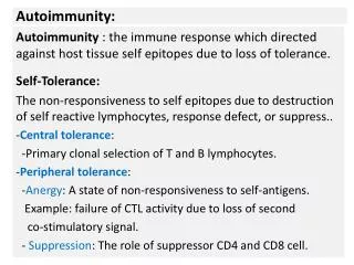
CORE 20. Autoimmunity and autoimmune diseases a. Origins of autoimmune diseases (1) Genetic factors: familial incidence; association with specific HLA (or MHC) haplotypes (2) Failure to maintain self-tolerance (either central or peripheral) (3) Loss of regulatory T cells (dysregulation of the cytokine network) (4) Expression of cryptic self epitopes (5) Inappropriate expression of MHC II molecules or co-receptors on specific tissue (6) Cross-reacting antigens and antigenic mimicry (7) Polyclonal B cell activation (8) Association of certain infectious diseases with the onset of autoimmunity (9) Hormonal influences
Pathogenic mechanisms of autoimmune diseases (see individual diseases) c. Examples of autoimmune diseases (prevalence, signs, symptoms, pathologic consequences of autoimmune mechanisms, examples of treatments [some key features of the diseases are given below])
Figure 11-17 How do we gain immune tolerance to self antigens? B cells: Autoreactive B cells are anergized or deleted in bone marrow, periphery and germinal centers in response to soluble antigens. T cells: Autoreactive T cells are deleted in the thymus. Autoreactive T cells are anergized or deleted in the periphery. Regulatory CD4+CD25+ T cells suppress autoreactive cells. Physical separation: separation of tissue antigens from immune cells. Limited expression of MHC II and B7 molecules
Figure 11-37 Autoimmune diseases result from defective self-tolerance. Requirement of T cell help in B cell responses
Influencing factors of autoimmune diseases • HLA (MHC) genotype/ Genetic background • Microbial infection: • Adjuvant effect: recruitment and activation of immune cells • Induction of MHC molecules and B7 • Molecular mimicry • Injury • Reveals cryptic auto-antigens from immune-privileged organs • Environmental factors and behavior • e.g. smoking and hygiene • Gender and sex hormones
Sympathetic ophthalmia: Physical trauma in one eye can initiate autoimmune response to both eyes • Eye anterior chamber is an immune-privileged site. Normally, auto-antigens in this site are not exposed to the immune system • Injury in one eye drain of eye proteins to the local lymph nodes immune responses on occasion, this causes blindness in the both damaged and undamaged eyes
Sex and autoimmunity Roles of sex hormones?
Figure 11-19 AutoImmune REgulator (AIRE) gene defect causes a wide range of autoimmune diseases Defective thymic deletion of autoreactive T cells due to inability to express peripheral antigens in the thymus. Autoimmune PolyEndocrynology Candidiasis Ectodermal Dystrophy Defective negative selection leads to autoimmunity
IPEX (immune dysregulation, polyendocrinopathy, enteropathy, X-linked syndrome) • Mutations in the T cell transcription regulator FOXP3 • FOXP3 is expressed by “regulatory T cells (Tregs)” • FOXP3 is essential for generation of Tregs. • Tregs suppress the activation of other immune cells.
Inflamed duodenum of IPEX patients IPEX patients don’t have FOXP3+ Tregs. Before After marrow transplantation Baud et al. NEJM, Volume 344:1758-1762 June 7, 2001 Number 23
Figure 11-23 Association of HLA allotype with autoimmune diseases: important!
Ankylosingspondylitis http://en.wikipedia.org/wiki/Image:Ankylosing_process.jpg
Figure 11-28 Food allergy Autoimmune disease Ciliac Disease: wheat flour gluten peptide-specific CD4+ T cells. HLA-DQ2/8 presents the peptide to CD4 T cells. CD4+ T cells activate macrophages and B cells. Development of anti-transglutaminase auto-antibodies. Destruction of villi structure
Infection plays an important role in causing autoimmunity. Molecular mimicry: antibodies to pathogens cross-react against host antigens. e.g. Rheumatic fever
Figure 11-31Figure 11-31 part 2 of 2 Molecular mimicry: similarity between pathogenic antigens and self-antigens causes the generation of auto-reactive T cells
Figure 11-32 Infection/inflammation induces IFN-g, which induces MHC class II expression on tissue cells and facilitates autoimmunity.
Figure 11-2 Autoimmune hemolytic anemia: type 2 Splenectomy is performed to reduce blood cell loss
type 2 Immune Thrombocytopenic Purpura (ITP) Antibodies attach to blood platelet, cells that help stop bleeding, and cause their destruction. Thrombocytopenia refers to decrease in blood platelet. Purpura refers to the purplish-looking areas of the skin and mucous membranes (such as the lining of the mouth) where bleeding has occurred as a result of decreased platelet.Some cases of ITP are caused by drugs, and others are associated with infection, pregnancy, or immune disorders such as systemic lupus erythematosus. About half of all cases are classified as "idiopathic," meaning the cause is unknown.
Goodpasture’s syndrome type 2 Autoantibodies IgG against the alpha3 chain of type IV collagen in the basement membranes throughout the body including lung and kidney (hemoptysis, dyspnea, anemia and nephritis) Renal Glomerulei is most sensitive to the antibody deposition and inflammatory response Treatments: plasma exchange and immunosuppressive drugs Infiltration with neutrophils and MNC
(2) Autoimmune Endocrine Diseases • Insulin-Dependent Diabetes Mellitus (Type II and IV; auto-antibodies to beta cell surface antigen, cytoplasmic antigen, and glutamic acid decarboxylase [associated with GABA synthesis]; HLA-DR proteins expressed on beta cells; HLA-DR3, HLA-DR4) • Chronic Thyroiditis (Hashimoto's Disease; Type II and IV; goiter; antithyroglobulin and antithyroid peroxidase are prevalent) • (c) Graves' disease (hyperthyroidism; anti-TSH receptor autoantibodies; MHC II [HLA-DR] expression on thyroid cells; HLA-DR3)
Autoimmune diseases of endocrine glands e.g. insulin- producing cells are attacked e.g. anti-insulin Ab Failure to produce adequate levels of cortisol.
Autoimmune diseases of endocrine glands: Thyroid gland Graves’ disease: Agonistic (stimulatory) autoantibodies against TSH receptor act as a ligand for the receptor (mimicking the natural TSH) leading to overproduction of thyroid hormone. Heat intolerance, nervousness, irritability, warm moist skin, weight loss, thyroid enlargement, bulging eyes. A Th2 type disease. Hyperthyroid disease. Treatment: thyroidectomy or destruction of thyroid by radioactive 131I
Figure 11-5 Graves’ disease
Autoantibodies can be passed from affected mothers to their new born babies: Graves’ disease
Figure 11-6 Hashimoto’s thyroiditis Antibodies and effector T cells specific for thyroid antigens are produced (attacking thyroid gland), leading to destruction of the thyroid gland and loss of thyroid hormone production. A Th1 type disease. Hypothyroid disease. Treatment: oral administration of thyroid hormone
IDDM:insulin-dependent diabetes mellitus Normal Insulitis Selective destruction of the beta cells of islets of Langerhans that produce insulin by the immune system (antibodies and T cells) Treatment: daily injection of insulin
(3) Autoimmune Liver and Gastrointestinal Diseases • Inflammatory Bowel Disease: Crohn's Disease (abnormality of mucosal T cell regulation; granulomatous reaction characteristic); Ulcerative colitis (Possibly Type II; continuous mucosal ulceration common) • Pernicious Anemia (antiparietal cell antibodies and anti-intrinsic factor antibodies) A chronic illness caused by impaired absorption of vitamin B-12 because of a lack of intrinsic factor (IF) in gastric secretions. VB-12 is a component of an enzyme required for thymine synthesis. (c) Autoimmune Chronic Active Hepatitis (HLA-B8/DR3; liver cells express MHC II proteins; anti-liver cell antibodies are present)
Autoimmune Rheumatic Diseases: Rheumatoid Arthritis (Type III and IV hypersensitive reactions; rheumatoid factors; vasculitis, synovitis; HLA-DR4; cellular and chemical effectors [PMN, TH1, TC, IL-1, TNF-alpha, IL-8, PGE2, LTB4) Systemic Lupus Erythematosus (Type II and III; multiple auto-antibodies; HLA-DR2, HLA-DR3; multiple organ involvement) Polymyositis/Dermatomyositis (HLA-DR3, HLA-DR; Type IV; serum muscle enzymes elevated)
Systemic Lupus (wolf) Erythematosus (skin rash) This facial rash is found in some SLE patients Affects 1 in 500 African or Asian women Abs to histone and DNA
SLE (Systemic Lupus Erythematosus): deposition of immune complexes • IgG against a wide variety of cellular constituents (e.g. nucleic acids). • Binding of antibodies to cell surface antigens causes inflammatory responses leading to cell and tissue destruction. • Immune complexes, deposited in blood vessels, kidney, joints and other tissues, causing tissue inflammation and destruction.
Figure 11-12 Rheumatoid arthritis: immune response to joints • Rheumatoid factor: IgM/G/A against the Fc region of IgG • Leukocyte infiltration in the joint synovium: CD4 T, CD8 T, B cells, neutrophils and macrophages • Plasma B cells produce rheumatoid factor • Inflammatory cells produce prostaglandins and leukotrienes, lysosomal enzymes, proteinases and collagenases • Treatment: anti-inflammatory and immunosuppressive drugs. e.g. anti-TNF-a
Smoking Injury generation of citruline residues activation of CD4 T cells RA
Autoimmune Neurologic Diseases (a) Multiple Sclerosis (abnormal T cell regulation; Type II and IV; inflammatory demyelination in CNS white matter resulting in "plaques"; auto-antibodies to MBP and PLP of myelin) (b) Acute Disseminated Encephalomyelitis (follows infection or vaccination; Type IV directed at MBP and other myelin antigens) (c) Acute Inflammatory Polyneuropathy (Guillain-Barre’ Syndrome; follows viral or Campylobacter infection; Type II and IV reaction to peripheral nerve antigens) (d) Myasthenia gravis (Type II autoimmune; Abs directed at acetylcholine receptors)
Guillain Barre’ Syndrome (acute idiopathic polyneuritis) • Guillain-Barrè (ghee-yan bah-ray) syndrome is a disorder in which the body's immune system attacks the peripheral nervous system. • The immune system starts to destroy the myelin sheath that surrounds the axons of many peripheral nerves • Initially, weakness or tingling sensations in the legs • Symptoms can increase in intensity until certain muscles cannot be used at all problems with breathing and heart beating • Plasmapheresis (to remove autoreactive Abs) and high-dose immunoglobulin therapy (mechanism unclear)