Download
1 / 23
230 likes | 385 Views
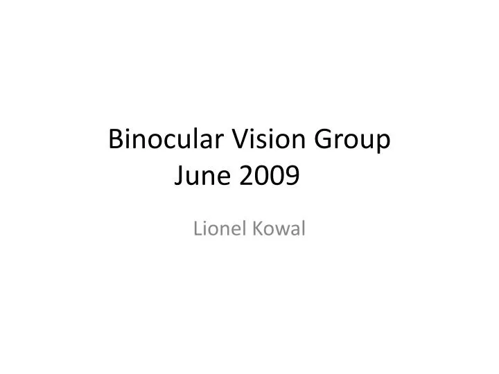
Binocular Vision Group June 2009 . Lionel Kowal. Bifocals in Down Syndrome Study (BiDS): design and baseline visual function. Nandakumar K, Leat SJ.School of Optometry, Waterloo, Ontario, Canada . . Optom Vis Sci. 2009 Mar;86(3):196-207.

E N D
Binocular Vision GroupJune 2009 Lionel Kowal
Bifocals in Down Syndrome Study (BiDS): design and baseline visual function.Nandakumar K, Leat SJ.School of Optometry, Waterloo, Ontario, Canada. • Optom Vis Sci. 2009 Mar;86(3):196-207. • PURPOSE: Among children and young people with Down syndrome (DS) there is a high prevalence of reduced accommodation. Prescribing bifocals for these patients has not become fully clinically accepted, although it would be anticipated to improve visual acuity (VA). The aim of this study is to investigate the impact of bifocal correction on VA, visual perceptual skills and early literacy development in children with DS who have reduced accommodation and who are provided with a bifocal correction • METHODS: We have chosen a longitudinal design with frequent measures of subtests of performance to control for progression with time. The main outcome measures are early literacy and visual perception skills. Secondary outcomes are VA and accommodative function. These are measured at baseline, the participant followed for 6 months when bifocals are prescribed if necessary, and the participants were followed for another 6 months with bifocals.
Bifocals in Down Syndrome Study (BiDS): design and baseline visual function. RESULTS n= 14 participants with DS aged 8 -19. At baseline 79% required a change in their distance spectacle prescription. ALL had reduced accommodation both before and after new single vision glasses were prescribed. None had an adverse reaction to 0.5 or 1% Cyclopentolate. All the subjects were able to perform either a distance or near crowded Patti-pics symbols test and 93% were able to perform both. There was a significant improvement of near VA with the new SV spectacles (p = 0.015). The mean binocular distance VA was 0.362 +/- 0.17 logMAR whereas binocular near VA was 0.489 +/- 0.235. • CONCLUSION: This study confirms previous findings of a high prevalence of reduced accommodation and shows that near VA is reduced compared to distance VA. The present results indicate that all subjects might benefit from bifocal
Bifocals and Down's syndrome: correction or treatment?Al-Bagdady M, Stewart RE, Watts P, Murphy PJ, Woodhouse JM. School of Optometry and Vision Sciences, Cardiff University, • Ophthalmic Physiol Opt. 2009 May 11. Purpose: Accommodation is reduced in approximately 75% of children with Down's syndrome (DS). Bifocals have been shown to be beneficial and they are currently prescribed regularly. Clinical observations suggest the likelihood of improving accommodative ability after bifocal wear. The aim of the study is to evaluate the potential use of bifocals as a treatment for the reduced accommodation. Methods: Clinical records of 40 children from the Cardiff Down's Syndrome Vision Research Unit, who were prescribed bifocals, were reviewed. Accommodation was noted before wearing the bifocals and during either their latest visit or when the children stopped using bifocals. Accommodation was reassessed during a follow up visit for the children who stopped wearing bifocals. Development of accommodation before bifocal commencement, age at bifocal prescription, gender, type of refractive error, visual acuity and the presence of strabismus were examined to evaluate their contribution to accommodation improvement.
Bifocals and Down's syndrome: correction or treatment? Results: The accommodative ability of 65% (n = 26) of the children improved (through the distance part of the lens) after using the bifocals. More than half of those developed accurate accommodation without the use of bifocals (n = 14). Accommodative responses did not show any improvement with age before the children began wearing bifocals. Accurate accommodation was sustained after returning to single vision lenses in all examined children. The age distribution of the children on bifocal commencement was diverse. Presence of strabismus, refractive error type, visual acuity and gender did not have any effect on gaining improvement. Conclusions: Bifocals are an effective correction for the reduced accommodation in children with DS and also act to improve accommodation with a success rate of 65%. Bifocal wear can therefore be temporary, i.e. a 'treatment' for the deficit, in at least one third of children
Bupivacaine injection of the lateral rectus muscle to treat esotropia.Scott AB, Miller JM, Shieh KR.The Smith-Kettlewell Eye Research Institute, San Francisco, California, USA. abs@ski.org • J AAPOS. 2009 April • PURPOSE: • We report results of a pilot trial of bupivacaine injection into extraocular muscles as a method of enlarging and strengthening the muscles to treat strabismus. • METHODS: • Bupivacaine, in volumes from 1.0 to 4.5 mL and concentrations from 0.75% to 3.0%, was injected into 1 lateral rectus muscle in each of 6 patients with comitant esotropia with the use of the electrical activity recorded from the needle tip to guide injection. Magnetic resonance imaging was performed before and at intervals after injection to estimate changes in muscle size. Clinical measures of alignment were made before and at intervals after injection. Two patients required a second injection for adequate effect.
Bupivacaine injection of the lateral rectus muscle to treat esotropia.Scott AB, Miller JM, Shieh KR.The Smith-Kettlewell Eye Research Institute, San Francisco, California, USA. abs@ski.org • Patient 3: preinjection, 25Δ esotropia (A); 15Δ esotropia before second injection, 161 days after first injection (B); and 11Δ esotropia on day 417 after second injection (C).
Axial MRI scans of Patient 5 injected with 1.0 mL of bupivacaine into the left lateral rectus: before injection (A) and 20 minutes after injection (B). Note the lack of bupivacaine in the important posterior third of the muscle.
Bupivacaine injection of the lateral rectus muscle to treat esotropia. RESULTS: • Four patients showed improved eye alignment, averaging 12(Delta), measured an average of 367 days after the last injection (range, 244-540 days). • Two patients were substantially unchanged. • Alignment improvement for all 6 patients averaged 8(Delta) (range, 0-14(Delta)). • Volumetric enlargement of the injected muscle, computed from magnetic resonance images, was 6.2% (range, -1.5% to 13.3%). • There was a positive correlation between alignment change and muscle enlargement averaging 0.65. Injection caused a retrobulbar hemorrhage in an unchanged patient that cleared without affecting vision. • CONCLUSIONS: • Bupivacaine injection improved eye alignment in 4 of 6 esotropic patients. There was a positive correlation between improved eye alignment and increased muscle size. Clinical and laboratory studies are underway to determine optimal dosages, effects in other strabismus conditions, and differential effects of bupivacaine on contractile and elastic muscle components
Long-term visual outcomes of laser-treated threshold retinopathy of prematurity: a study of refractive status at 7 years.Yang CS, Wang AG, Sung CS, Hsu WM, Lee FL, Lee SM Taiwan • Eye. 2009 Apr 3. Purpose • To assess the long-term visual outcomes and refractive status in patients with diode laser-treated threshold retinopathy of prematurity (ROP), and to investigate the causes of impaired visual function. Method • 60 eyes of 30 consecutive patients with diode laser-treated threshold ROP were recalled for assessment at the age of 7 years or more .
ResultsLong-term visual outcomes of laser-treated threshold retinopathy of prematurity: a study of refractive status at 7y • 38 eyes (65.5%) : 6/12 or better vision, • unfavourable visual outcome (6/60 or worse) occurred in four eyes (6.9%). • One eye (1.7%) had unfavourable structural outcome. • 46 eyes (77%) were myopic, overall mean spherical equivalent was -3.9 D. • Anisometropia (>/=1.5 D) in 14 patients (46.7%). • Strabismus in 9 patients (30%). • intraventricular haemorrhage (IVH) were identified in 8 children (26.7%), • periventricular leucomalacia (PVL) in 8 children (26.7%) • cerebral palsy (CP) in 4 children (13.3%). • statistically significant association of strabismus with PVL (P=0.002). • anisometropia was a significant risk factor associated with poor visual outcome of 6/15 or worse in laser-treated ROP (P=0.002).
Long-term visual outcomes of laser-treated threshold retinopathy of prematurity: a study of refractive status at 7y Conclusion • The majority of patients with diode laser-treated threshold ROP had favourable anatomical and visual outcomes. However, anisometropia, advanced refractive error, strabismus, and perinatal neurological events remain important causes of impaired visual function. Long-term follow-up is very important for early detection and timely treatment of these ocular morbidities.
Spontaneous consecutive exotropia in childhoodSenior JD, Chandna A, O'Connor AR .Liverpool, UK. • Strabismus. 2009 Jan-Mar;17(1):33-6.. • PURPOSE: • Consecutive exotropia is commonly the result of a surgical correction of a primary esotropia but may develop spontaneously. This study examines the clinical characteristics of patients who have developed a spontaneous consecutive exotropia and relates the key features to previously reported findings. • METHODS: • Patients were identified by routine clinical practice from May 2000 to December 2003 by retrospective case note analysis. Patients that presented with an esotropia that became an exotropia (without surgery or botulinum toxin A) were included.
Spontaneous consecutive exotropia in childhood • RESULTS: • This study consisted of 14 patients who presented with an esotropia from early weeks of life to almost 4 years. • No patients were undergoing occlusion treatment at the onset of the exotropia and no subject had dense amblyopia. • The most common characteristics included • (1) age of onset of esotropia of 24 months or less; • (2) hypermetropic refractive error of +5 DS mean spherical equivalent; • (3) lack of binocular single vision. • CONCLUSION: • The results highlight the variability of the condition with no single consistent finding, with the lack of binocular single vision, early onset esotropia, and high hypermetropia being common findings. Amblyopia was not an important characteristic
LK case > When age 22 mo, I saw this girl with intermittent ET.> Had been admitted age 11w with upbeat nystagmus and upgaze palsy. Normal> MRI.> At age 22mo, there were a few beats of high frequency small amplitude> vertical nystagmus every now and then [?as described in CSNB by Gottlob in> BJO 2008].> Straight for distance, intermittent near ET'. Cyclo +0.5 DSOU.> Age 26 mo: infrequent transient ET and alternating height seen by me. No> nystagmus seen. Parents mention nyctalopia; ERG suggests CSNB.>> And NOW: age 35 mo: intermittent XT> Constant for Distance 30^; straight for N.> Was given -2 DSOU @ St elsewhere's.> 6/10 OU with Allen Pix.>> Apparent spontaneous consec XT [from Et to XT] is pretty unusual.> Anyone think the CSNB is a factor?>> Thanks>>> Dr Lionel KOWAL>
LK case Dear Lionel,I have done strab surgery on a few CSNB patients. All very difficult to keep straight. A common pattern is that they are straight for a few weeks to months and then develop some form of strab. One patient I am currently treating will go from a pure horizontal deviation to a vertical deviation after surgery or the reverse. I have attributed this to the weak peripheral visual field secondary to the retinal dystrophy.Secondly, you can expect her to become a myope, even possibly a severe myope, with time. So over-minussing at this age would be consistent with her future course.All the best, Sherwin isenberg, UCLA>
A novel experimental method for measuring vergence and accommodation responses to the main near visual cues in typical and atypical groupsHorwood AM, Riddell PM. School of Psychology and Clinical Language Sciences, University of Reading, Reading, UK. a.m.horwood@reading.ac.uk • Strabismus. 2009 Jan-Mar;17(1):9-15. Links. • Binocular disparity, blur, and proximal cues drive convergence and accommodation. • Disparity is considered to be the main vergence cue and blur the main accommodation cue. • We have developed a remote haploscopic photorefractor to measure simultaneous vergence and accommodation objectively in a wide range of participants of all ages while fixating targets at between 0.3 and 2 m. By separating the three main near cues, we can explore their relative weighting in three-, two-, one-, and zero-cue conditions. • Disparity can be manipulated by remote occlusion; blur cues manipulated by using either a Gabor patch or a detailed picture target; looming cues by either scaling or not scaling target size with distance. • In normal orthophoric, emmetropic, symptom-free, naive visually mature participants, disparity was by far the most significant cue to both vergence and accommodation. Accommodation responses dropped dramatically if disparity was not available. Blur only had a clinically significant effect when disparity was absent. Proximity had very little effect. There was considerable interparticipant variation. We predict that relative weighting of near cue use is likely to vary between clinical groups and present some individual cases as examples. We are using this naturalistic tool to research strabismus, vergence and accommodation development, and emmetropization
A novel experimental method for measuring vergence and accommodation responses to the main near visual cues in typical and atypical groups • In normal orthophoric, emmetropic, symptom-free, naive visually mature participants, disparity was by far the most significant cue to both vergence and accommodation. • Accommodation responses dropped dramatically if disparity was not available. • Blur only had a clinically significant effect when disparity was absent. • Proximity had very little effect. • There was considerable interparticipant variation. • We predict that relative weighting of near cue use is likely to vary between clinical groups and present some individual cases as examples. We are using this naturalistic tool to research strabismus, vergence and accommodation development, and emmetropization
The influence of head tilt on ocular torsion in patients with superior oblique muscle palsy Burton J. Kushner, MD • JAAPOS Volume 13, Issue 2, Pages 132-135 (April 2009) • Introduction • Patients with superior oblique muscle palsy often assume a compensatory head tilt to the contralateral side of the affected eye. This tilt serves to decrease the hypertropia and, in theory, might decrease the excyclotropia. • Methods • A prospective investigation was undertaken of the effect of forced head tilt to the right and left on torsion and the hypertropia in patients with unilateral superior oblique muscle palsy.
The influence of head tilt on ocular torsion in patients with superior oblique muscle palsy Burton J. Kushner, MD • The Cervical Range of Motion (CROM) device was used to measure the spontaneous compensatory head tilt in all patients while they read Snellen optotypes at threshold acuity at a distance of 6 meters while wearing appropriate optical correction. • The angle of misalignment was measured in the nine diagnostic fields of gaze at 6 meters and on head tilt right and left at 6 meters with the appropriate optical correction in place.
The influence of head tilt on ocular torsion in patients with superior oblique muscle palsy Burton J. Kushner, MD • Subjective torsion was measured with the double Maddox rod test using white and red lenses that were etched to accurately denote the 90° and 180° positions. First, the paretic eye was occluded and the white Maddox rod was placed over the nonparetic eye. The Maddox rod was then rotated until the patient perceived the white line as being vertical while the head was in the straight upright position. Next the paretic eye was uncovered and the red Maddox rod was placed in front of it, initially oriented so that the line was approximately 30° counterclockwise from the vertical position. It was then slowly rotated clockwise until the patient perceived the red and white lines as being parallel or superimposed, and the amount of torsion was recorded to the nearest 1°. The red Maddox rod was then rotated approximately 30° clockwise from the vertical position and slowly rotated back toward the 90° orientation until the subject saw the lines as parallel or superimposed, and this reading of torsion also was recorded. • These were repeated in sequence for a total of 6 readings, 3 from each direction to assess accuracy and reproducibility for each subject. • The 6 readings were then averaged, rounded to the nearest 1°, and used for the data analysis. • If any of the readings varied by more than 2° for any subject, the patient was considered unreliable and excluded from the study.
The influence of head tilt on ocular torsion in patients with superior oblique muscle palsy Burton J. Kushner, MD • This patient has a left superior oblique muscle palsy associated with a 10° left excyclotropia in the head-erect position (A). With a head tilt of approximately 30° to the right, he orients the white Maddox rod over the nonparetic right eye as being vertical when it is positioned at 55° in the trial frame (B). The paretic left eye perceives the lines as parallel when the red Maddox rod is oriented at 45°, indicating 10° of excyclotropia. Note that there is a difference of 5° between the 30° of head tilt and the 35° of rotation needed to perceive the white Maddox rod as vertical. This probably represents partial compensatory countertorsion
The influence of head tilt on ocular torsion in patients with SOP Results • 20 patients with unilateral SOP had a mean decrease in their hypertropia of 6.4Δ ± 3.9Δ on forced head tilt from the head-erect position toward the contralateral side. This finding represented a decrease of 62% of the hypertropia found in the head-erect position. The difference was significant (p < 0.001, paired t-test). • The mean decrease of the excyclotropia was only 0.25° ± 0.6° on contralateral head tilt, which represented 3% of the excyclotropia in the head-erect position. This difference was not significant (p < 0.06). • The percent by which the hypertropia decreased from the head-erect position to that found on contralateral head tilt was significantly greater than the amount by which the excyclotropia decreased (p < 0.001). • Conclusions • With contralateral head tilt, patients with superior oblique muscle palsy demonstrate a significant decrease in their hypertropia but essentially no change in their excyclotropia. The compensatory head tilt they manifest appears to serve the purpose of decreasing the vertical but not the torsional misalignment