Download

1 / 23
390 likes | 2.88k Views
Preparation of Blood films (Making, Fixing & Staining technique). Dr Maliha Sumbul. SMEARS USING BLOOD ANTICOAGULATED WITH EDTA SHOULD BE MADE WITHIN 2-3 HOURS OF BLOOD COLLECTION. MAKING. Types. Cover glass smear Wedge smear Spun smear Buffy coat smear Thick blood smear.

E N D
Preparation of Blood films(Making, Fixing & Staining technique) Dr Maliha Sumbul
SMEARS USING BLOOD ANTICOAGULATED WITH EDTA SHOULD BE MADE WITHIN 2-3 HOURS OF BLOOD COLLECTION
Types • Cover glass smear • Wedge smear • Spun smear • Buffy coat smear • Thick blood smear
The cover glass smear • More even white cells distribution • More time consuming • Difficult technique • Cover glasses too small for most automated stainers • Harder to label • Easily broken METHOD
Wedge blood smears METHOD (HEAD, BODY AND TAIL) IDEAL SMEAR POOR SMEAR
The Automated Spun smear • Uses more easily handled and labeled glasss slide • Even distribution of white blood cells • Red cells free of distortion • Method: • Hemaspinner • Slide in a platen • Thin monolayer of cells
The buffy coat smear • When pt’s WBCs is < 1.0 x 10⁹ / L – FOR DIFFERENTIAL COUNT • This procedure concentrates the nucleated cells present in the blood • Wintrobe tube filled with capillary pipet • C/G for 15 min at 1500 G • Remove plasma • Pipet out buffy coat with small amount of red cells on a glass slide – mix well • Prepare wedge smears or cover glass smears on a separate slide
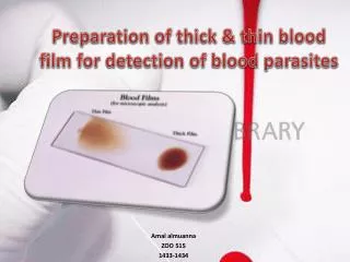
Thick blood smears • Used for blood parasites, for example, MALARIA METHOD
Characteristics of a Good Smear • 1) Thick at one end, thinning out to a smooth rounded feather edge. • 2) Should occupy 2/3 of the total slide area. • 3) Should not touch any edge of the slide. • 4) Should be margin free, except for point of application.
Adjustment of the Smear Length • Increasing the angle of the spreader slide will decrease the length of the smear. Decreasing the angle will increase the smear length.
FIXING (METHANOL)
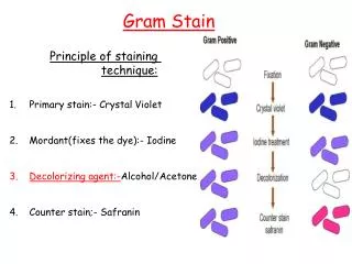
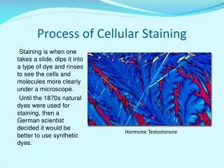
ROMANOWSKY STAINING • Universally used for staining blood films • Polychromatic stains – dyes produce multiple colours • Main components: - AZURE B or Trimethylthionin (oxidation product of methylene blue) - positively charged (cationic/basic dye) - binds to acid structures such as nucleic acids – DNA/RNA - BLUE/PURPLE COLOUR - Eosin Y - negatively charged (anionic/acidic dye) - stains the basic components of the cells ( Hb, eosinophil granules) - ORANGE/PINK COLOUR
Examples • LEISHMAN’S • WRIGHT’S • GIEMSA • FIELD STAIN – rapid stain
Preparation of Leishman stain • 1.5 grams L powder • 1 litre - Methanol MATURATION TIME
BUFFER SOLUTION(Sorenson’s phosphate buffer) • After adding the stain, a buffer solution is added • Ionization occurs, during which time staining takes place ( a physical process of converting an atom or molecule into an ion by adding or removing charged particles such as electrons or other ions ) • STOCK A – KH2 PO4 (anhydrous monobasic potassium phosphate) 9.1 gms + 1 litre D/W • STOCK B – Na2HPO4 (anhydrous dibasic sodium phosphate) 9.5 gms + 1 litre D/W
STAINING PROCEDURE FOR LS • Make slide and air dry • Place air dried slide on a level staining rack with smear side up • Pour LS and leave for 1 min • Pour buffer 1: 2 ratio • Gentle blowing to mix – metallic green sheen – 10 min • Wash with tap water • Wipe the back with cotton to remove stain • Air dry
POINTS TO REMEMBER • pH by phosphate buffer • Staining rack – leveled • Insufficient washing • Excessive rinsing • To restain – remove the original stain with methanol • Stock buffer solution must be prepared fresh each week • Daily check pH with filter paper