Download
1 / 30
310 likes | 721 Views
OA 12.4. What is the following structure:. Chapter 18 (pp 471-486 ). T he Knee. Anatomy. O bjectives. Identify… The bones of the knee The ligaments of the knee The muscles of the knee The tendons of the knee The blood vessels & nerves of the knee Other structures. Bones. Femur (1)

E N D
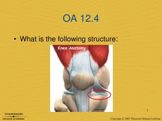
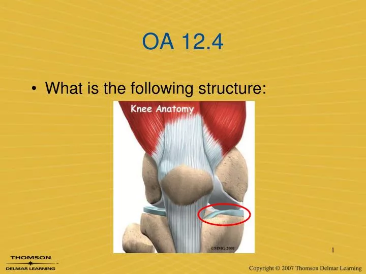
OA 12.4 • What is the following structure:
Chapter 18 (pp 471-486) The Knee
Objectives Identify… • The bones of the knee • The ligaments of the knee • The muscles of the knee • The tendons of the knee • The blood vessels & nerves of the knee • Other structures
Bones • Femur (1) • Tibia (3) • Fibula (4) • Patella (2)
Bones: landmarks • The proximal end of the tibia is called the plateau • Attachment site for ACL, PCL, Meniscus • The proximal end of the fibula is called the head (apex) • Attachment site for LCL
Bones: landmarks • The tibial tuberosity is the bony outgrowth on the anterior aspect of the tibia • Attachment site for patellar tendon
Bones: landmarks • The distal end of the femur is has two condyles, and just superior to those are epicondyles • Medial = MCL & lateral = LCL
Bones • The patella is the largest sesamoidbone in the body • Housed within quadriceps/ patellar tendon
Menisci • The knee contains two meniscus– medial & lateral • Medial = C shaped • Lateral = O shaped
Menisci • The meniscus is only partly vascularized (only part receives blood flow) • Known as the vascularor avascular zone
Menisci • Fibrocartilage • Deepens the joint • Increases stability • Absorbs shock • Lubricates the joint
Ligaments • There are four main stabilizing ligaments in the knee • Two cruciates (crossing) • Two collaterals(on the sides) • Anterior cruciate ligament • Posterior cruciate ligament • Medial collateral ligament • Lateral collateral ligament
Ligaments ACL PCL Posterior-lateral tibia to medial condyle of femur Prevents internal rotation of the tibia & guides the knee during flexion • Anterior-medial tibia to lateral condyle of femur • Three bands • Prevents internal rotation of the tibia & anterior translation
Ligaments MCL LCL Lateral epicondyle of the femur to head of the fibula Protects against varusforces • Medial epicondyle of femur to tibia • Protects the knee from valgusforces & external rotation of the tibia
Articulations • The knee is comprised of three articulations • Tibiofemoral= tibia & femur • Tibiofibular= proximal tibia & fibula • Patellofemoral= patella & femur
Muscles & tendons • Anterior aspect – extend the knee (and flex the hip) • Quadriceps femoris group • Vastusmedialis • Vastusintermedius • Vastuslateralis • Rectus femoris • Sartorius*
Muscles & tendons • The quadriceps group forms the quadriceps tendon • Pulls on the patella to extend the knee
Muscles & tendons • Posterior aspect – flex the knee* (and extend the hip) • Hamstrings group • Semitendinosis • Senimembranosis • Biceps femoris • Gastrocnemius • Plantaris • Popliteus
Muscles & tendons • Three muscles join to form the pes anserine group, which insert on the anterior tibia • Sartorius, Gracilis, SemiTendinosus • Acronym: SGT. (sargeant) • Knee flexors
Muscles & tendons • The lateral aspect of the knee is controlled/stabilized by the iliotibial band (IT Band (8)) • Connective tissue that attaches the TFL muscle to the knee at Gerdy’s Tubercle (7)
Other structures • The posterior aspect of the knee is known as the popliteal fossa • Popliteus muscle • Popliteal tendon • Popliteal artery • Popliteal nerve
Other structures • Bursae – as many as two dozen around the knee • Suprapatellar (A&B) • Prepatellar (C&D) • Infrapatellar(F, G, H) • Pes anserine (I)
Other structures • Fat pads – exist to cushion the knee
Other structures • Nerve supply • Tibial nerve • Common peroneal nerve • Femoral nerve • Located on… • Posterior aspect • Lateral aspect • Anterior-Medial aspect
Other structures • Blood supply • Femoral artery (2) popliteal artery (3) • Pulse is felt at the popliteal artery, in the popliteal fossa