Download
1 / 7
70 likes | 177 Views

E N D
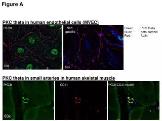
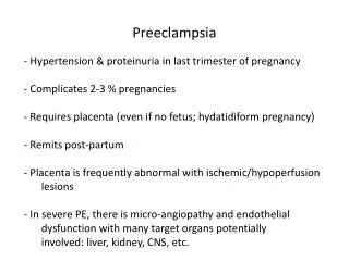
Glutamine reduces ICAM-1 expression in endothelial cells activated by preeclampsiaChun-Sen Hsu, MD,a Szu-Yuan Chou, MD,a Wan-Chun Chiu, MS,b So-Jung Liang, MD, a Chih-Fen Wu, MD,a Chiu-Li Yeh, MS,b Sung-Ling Yeh, PhDbDepartment of Obstetrics and Gynecology, Taipei Medical University-Municipal Wan-Fang Hospitala, and Institute of Nutrition and Health Sciences, Taipei Medical Universityb, Taipei, Taiwan • This study was supported by research grant 93TMU-WFH-10 from Taipei Medical University-Municipal Wan-Fang Hospital, Taipei, Taiwan • Corresponding author: • Sung-Ling Yeh, PhD • School of Nutrition and Health Sciences • Taipei Medical University • 250 Wu-Hsing Street, Taipei, Taiwan 110, ROC • Tel: 8862-27361661 ext. 6551-115 • E-mail: sangling@tmu.edu.tw • Condensation • Preeclamptic women have lower plasma glutamine levels than normal pregnant women. Glutamine administration at levels similar to physiologic conditions reduces endothelial cell ICAM-1 expression stimulated by preeclamptic plasma.
Glutamine reduces ICAM-1 expression in endothelial cells activated by preeclampsia • Chun-Sen Hsu, MD,a Szu-Yuan Chou, MD,a Wan-Chun Chiu, MS,b So-Jung Liang, MD, a Chih-Fen Wu, MD,a Chiu-Li Yeh, MS,b Sung-Ling Yeh, PhDb • OBJECTIVE: Our purpose was to investigate whether plasma glutamine (GLN) was depleted in women with preeclampsia, and whether GLN administration comparable to physiologic concentrations can decrease cellular adhesion molecule expression in human umbilical vein endothelial cells (HUVECs) induced by plasma from preeclamptic women. • STUDY DESIGN: Blood samples collected from 20 women with preeclampsia and 10 normal pregnant women were assayed for plasma GLN levels. HUVECs were cultured in medium-199 containing fetal calf serum, antibiotics, growth factor, and different concentrations (0, 300, 500, and 1000 uM) of GLN for 24 h. We stimulated these cells for up to 6 h with sera from patients with preeclampsia and then determined the expressions of the intercellular cell adhesion molecule (ICAM)-1 and vascular cell adhesion molecule (VCAM)-1 in endothelial cells by flow cytometry. • RESULTS: Women with preeclampsia exhibited significantly lower plasma GLN concentrations compared to normal pregnant women. There were no differences in VCAM-1 expression; however, ICAM-1 expression in HUVECs was significantly lower in the 500 uM GLN group than the 0, 300, and 1000 uM groups at 3, 4.5 and 6h. • CONCLUSIONS: This study revealed that plasma GLN levels from women with preeclampsia were significantly lower than those of normal pregnant women. GLN administration at levels similar to physiologic conditions reduces HUVEC ICAM-1 expression stimulated by preeclamptic plasma. • KEY WORDS: Preeclampsia, Glutamine, Intercellular cell adhesion molecule, Vascular cell adhesion molecule
Endothelial activation/dysfunction is a central pathogenic feature in women with preeclampsia, which is a pregnancy-specific syndrome with multiple system disorders.1,2 The mechanisms leading to this dysfunction have not been fully clarified. Previous reports revealed that cell adhesion molecules (CAMs) are increased in the serum of patients with preeclampsia, and soluble CAM levels are associated with the severity of the disease.3,4 Adhesion molecules play a key role in cell-cell interactions and cell-extracellular matrix interactions. On activated endothelium, intracellular adhesion molecule 1 (ICAM-1) and vascular cell adhesion molecule 1 (VCAM-1) are expressed. ICAM-1 and VCAM-1 are important for the adhesion of leukocytes to activated endothelium.5,6 Previous reports showed that ICAM-1 and VCAM-1 expression in human umbilical endothelial cells (HUVECs) was induced by plasma from preeclamptic patients.7,8 • Glutamine (GLN) is a critical substrate for enterocytes and rapidly proliferating immune cells. Several studies have demonstrated that GLN has immuno-enhancing properties.9-11 Previous reports have revealed that a relatively GLN-deficient state is created by the catabolic process, and GLN supplementation can correct this nutritional deficiency and hence improve outcomes.12,13 GLN is considered an essential amino acid during certain inflammatory conditions.14,15 A study by Fukatsu et al.16 showed that compared with conventional total parenteral nutrition, GLN-supplemented parenteral nutrition reduced ICAM-1 expression in intestinal homogenates. Also, Arndt et al.17 demonstrated that GLN administration reduced leukocyte adhesion and transmigration in intestinal inflammation in rats. As far as we know, there is no study investigating the effect of GLN on the expression of CAM in preeclampsia. We hypothesized that chronic inflammation during preeclampsia results in depletion of plasma GLN, and GLN administration comparable to physiologic concentration may decrease CAM expression in HUVECs induced by plasma from preeclamptic women.
Materials and Methods • Patients and sample information • The pregnant women were recruited from Taipei Medical University-Municipal Wan-Fang Hospital, Taipei, Taiwan. Women with singleton pregnancies and no chronic disease were included in this study. Normal pregnancy (n = 10) was one in which the women remained normotensive and non-proteinuric and had no medical complications. Preeclampsia (n = 20) was diagnosed as a blood pressure of at least > 140/90 mmHg on at least two occasions occurring after the 20th week of gestation accompanied by more than 300 mg protein in a 24-h urine collection or 1+ proteinuria detected on a reagent strip on two occasions more than 4 h apart.4 The mean gestational age at the time of blood sampling was 37.6 0.9 weeks for the controls and 37.3 1.9 weeks for the preeclamptic women. Among the 20 preeclamptic patients, we collected 5 plasma samples from women with severe preeclampsia for an in vitro study. Severe preeclampsia was defined as a blood pressure > 160/110 mmHg on two occasions 6 h apart and proteinuria corresponding to > 2+ detected on a reagent strip on two occasions more than 4 h apart.4 The protocol was approved by the Wan-Fang Hospital Ethical Committee, and all subjects provided written informed consent prior to their participation. • HUVEC isolation and culture • HUVECs were isolated as previously described.18 Briefly, the umbilical vein was cannulated and washed with PBS and then perfused with PBS containing 0.1% collagenase for 10 min at 37 ℃ in 5% CO2. Primary cells were collected and plated into 75-cm2 tissue culture flasks with medium-199 (M-199) containing 20% fetal calf serum (FBS), 20 mM NaHCO3, 25 mM HEPES, antibiotics (100 U/ml penicillin and 100 ug/ml streptomycin), 10 IU/ml heparin sodium, and 15mg/L endothelial cell growth factor at 37 ℃ in a 5% CO2 /95% humidity atmosphere. Cells were serially passaged 2-3 times for the experimental assay. HUVECs (1 × 10⁵ cells/well) from second subcultures were grown on 24-well plates. Every 48-72 h, the medium was replaced until the cells reached confluence, and then they were detached using typsin-EDTA. When cells reached a confluent monolayer, they were then incubated in M-199 (with 20% FBS) with different concentrations of GLN (0, 300, 500, and 1000 uM) for 24 h. Subsequently, cells were washed twice with PBS, cultured with various concentrations of GLN (without FBS), and stimulated with 10% plasma from severe preeclamptic women. Analytic flow cytometry analysis was then performed for ICAM-1 and VCAM-1 expression.
Measurements and analytical procedures • Plasma amino acid analysis • Antecubital venous blood was drawn from fasting women into EDTA-containing tubes. Plasma amino acids were analyzed by standard ninhydrin technology (Beckman Instruments, model 6300, Palo Alto, CA, USA), after deproteinization of the plasma with 5% salicylic acid.19 • ICAM-1 and VCAM-1 expression of HUVECs • HUVEC surface expression of ICAM-1 and VCAM-1 was measured after stimulation by preeclamptic plasma for 1.5, 3, 4.5, and 6 h, because CAM expression reached a peak at 6 h after stimulation. After removing the supernatant, cells were washed twice with PBS, incubated with 100 ul of M-199 (FBS free and containing 5 mM EDTA), and incubated for a further 30 min at 4 ℃ with fluorescein-conjugated mouse anti-human VCAM-1 (CD 106) and phycoerythrin-conjugated mouse anti-human ICAM-1 (CD 54). The fluorescence intensity of a 5000-cell population was counted and analyzed by flow cytometry (Coulter, Miami, FL, USA). • Measurement of IL-8 and NO in culture medium • The cell suspension was collected, and IL-8 concentrations were determined by an enzyme-linked immunosorbent assay (BioSource International, Camarillo, CA, USA). NO concentrations were determined with a commercial kit (R&D Systems, Minneapolis, MN, USA). • Statistical analysis • Data are expressed as the mean SD. Differences among groups were analyzed by ANOVA using Duncan’s test. A p value of < 0.05 was considered statistically significant. • Results • Plasma GLN levels • Plasma GLN levels in normal pregnant women were significantly higher than those in the preeclampsia groups (509.5 52.1 vs. 349.8 55.1 umol/L, p < 0.05). • CAM expression in HUVECs induced by plasma from preeclamptic women • There were no differences in VCAM-1 expression in HUVECs among various GLN concentrations at each time point (Fig. 1). However, ICAM-1 expression in HUVECs was significantly lower in the 500 uM GLN group than in the 0, 300, and 1000 uM groups at 3, 4.5, and 6 h (Fig. 2). • IL-8 and NO production from endothelial cells • There were no differences in IL-8 (Fig. 3) and NO production (Fig. 4) among various GLN concentrations at different time points after incubation.
Comment • This study is the first to report that plasma from women with preeclampsia had significantly lower GLN levels than that of normal pregnant women; this finding is compatible with previous reports that plasma GLN is reduced during catabolic conditions.12-14 Therefore, we pretreated endothelial cells with low (300 uM), approximately physiological (500 uM) and high (1000 uM) GLN levels to investigate whether physiological levels of GLN may have an effect on reducing CAM expression in endothelial cells stimulated by preeclamptic plasma. Our study demonstrated that GLN administration at levels similar to physiological conditions reduced ICAM-1 expression in vascular endothelial cells. Since ICAM-1 is the ligand counterpart of integrins on polymorphonuclear neutrophils, reduced ICAM-1 expression may decrease polymorphonuclear neutrophil-endothelium interactions and thus attenuate tissue injury.5 This finding may provide information that is clinically useful for managing pregnant women with preeclampsia. • It has been suggested that preeclampsia may represent an excessive maternal inflammatory response to pregnancy,20 and some cytokines are thought to be associated with the initiation of preeclampsia.21,22 IL-8 is a potent chemoattractant for leukocytes, initiates the acute inflammatory cascade, and is an early marker of the inflammatory process. A previous report revealed that an increase in IL-8 may result in increased endothelial permeability.22 A study by Fowler et al.23 showed that exogenous NO suppresses IL-8 gene expression in activated endothelial cells by inhibiting NF-κB binding to DNA. Since ICAM-1 is regulated transcriptionally by NF-κB, inhibition of NF-κB may result in reduced ICAM-1 expression.8 An in vitro study by Huang et al.24 showed that GLN decreases LPS-induced IL-8 production in Caco-2 cells. In this study, no differences in IL-8 and NO levels between various GLN concentrations at different time points were observed. This finding may indicate that the effect of GLN on reducing endothelial ICAM-1 expression might not have an association with IL-8 and NO production. These results are consistent with reports of others, those studies showed that compared with normal pregnant women, maternal plasma from preeclamptic women does not affect endothelial cell NO synthase mRNA expression or IL-8 production.22,25 It is possible that other factors could be sources of endothelial activation in the maternal circulation during preeclampsia. • A study by Hong et al.26 revealed that GLN-supplemented nutrition protects the liver during hepatic injury by preserving glutathione stores. An in vitro study by Babu et al.27 also found that GLN reverses the beneficial effect in preventing liver damage possibly mediated via GSH synthesis. GSH is a major antioxidant and a vital component of a host’s defense. GLN was found to be rate limiting for GSH synthesis, and the availability of GLN is critical for the generation of GSH stores.28 A previous study showed that plasma from women with preeclampsia had significantly higher malondialdehyde and lipid peroxide levels than those of normal pregnant women.6 Lipid peroxide and oxidative stress upregulate endothelial NF-κB activity and ICAM-1 expression.6 Determining whether administration of physiological levels of GLN reduces oxidative stress and thus decreases ICAM-1 expression requires further investigation. • In summary, this study revealed that plasma from women with preeclampsia had significantly lower GLN levels compared to that of normal pregnant women. GLN administration at levels similar to physiologic conditions reduces HUVEC ICAM-1 expression stimulated by preeclamptic plasma.
References: • Roberts JM, Taylor RN, Musci TJ, Rodgers GM, Hubel CA, Mclaughlin MK. Preeclampsia: an endothelial cell disorder. Am J Obstet Gynecol 1989;161:1200-4. • Wang Y, Adair CD, Coe L, Weeks JW, Lewis DF, Alexander JS. Activation of endothelial cells in preeclampsia: increased neutrophil-endothelial adhesion correlates with up-regulation of adhesion molecule P-selectin in human umbilical vein endothelial cells isolated from preeclampsia. J Soc Gynecol Invest 1998;5:237-43. • Coata G, Pennacchi L, Bini V, Liotta L, Di Renzo GC. Soluble adhesion molecules: marker of preeclampsia and intrauterine growth restriction. J Matern Fetal Neont Med 2002;12:28-34. • Djurovic S, Schjetlein R, Wisloff F, Haugen G, Berg K. Increased levels of intercellular adhesion molecules and vascular cell adhesion molecules in preeclampsia. Br J Obstet Gynecol 1999;104:466-70. • Carlos T, Harian J. Leukocyte-endothelial adhesion molecules. Blood 1994;54:2068-101. • Jang Y, Linoff AM, Plow EF, Topol EJ. Cell adhesion molecules in coronary artery disease. J Am Coll Cardiol 1994;24:1591-601. • Heyl W, Handt S, Reister F, Gehlen J, Mittermayer C, Rath W. The role of soluble adhesion molecules in evaluating endothelial cell activation in preeclampsia. Am J Obstet Gynecol 1999;180:68-72 • Takacs P, Kauma SW, Sholley MM, Walsh SW, Dinsmoor MJ. Increased circulating lipid peroxides in severe preeclampsia activate NF-B and upregulate ICAM-1 in vascular endothelial cells. FASEB 2001;15:279-81. • Calder PC. Glutamine and the immune system. Clin Nutr 1994;13:2-8. • Heberer M, Babst R, Juretic A, Gross T, Horig H, Harder F, et al. Role of glutamine in the immune response in critical illness. Nutrition 1996;12(Suppl):S71-2. • Wilmore DW, Shabert JK. Role of glutamine in immunologic responses. Nutrition1998;14:618-26. • Hammerquist F, Wernerman J, Al R, Vonder D, Vinners E. Addition of glutamine to total parenteral nutrition after elective abdominal surgery spares glutamine in muscle, counteracts the fall in muscle protein synthesis and improves nitrogen balance. Ann Surg 1989;209:455-61. • Gianotti L, Alexander JW, Gennari R, Pyles T, Babcock GF. Oral glutamine decreases bacterial translocation and improves survival in experimental gut-origin sepsis. J Parenter Enter Nutr 1995;19:69-74. • Lacey JM, Wilmore DW. Is glutamine a conditionally essential amino acid? Nutr Rev1990;48:297-309. • Willmore DW. The effects of glutamine supplementation in patients following elective surgery and accidental injury. J Nutr 2001;131:2543S-9S. • Fukatsu K, Lundberg AH, Kudsk KA, Hanna MK and Zarzaur BL. Modulation of organ ICAM-1 expression during IV-TPN with glutamine. Shock 2001;15:24-8. • Arndt H, Kullmann F, Reuss F, Scholmerich J and Palitzsch KD. Glutamine attenuates leukocyte-endothelial cell adhesion in indomethacin-induced intestinal inflammation in the rat. J Parenter Enter Nutr 1999;23:12-8. • Jaffe EA, Nachman RL, Becker CG, Minick RC. Culture of human endothelial cells derived from umbilical veins. Clin Invest 1973;52:2745-56. • Smith RJ, Panico K. Automated analysis of o-phthalaldehyde derivatives of amino acids in physiological fluids of reverse phase high performance liquid chromatography. J Liq Chromatogr 1985;8:1783-95. • Redman CW, Sacks GP, Sargent IL. Preeclampsia: an excessive maternal inflammatory response to pregnancy. Am J Obstet Gynecol 1999;180:499-506. • Ellis J, Wennerholm UB, Bengtsson A, Lilja H, Petterssson A, Sultan B, et al. Levels of dimethylarginines and cytokines in mild and severe preeclampsia. Acta Obstet Gynecol Scand 2110;80:602-8. • Wang Y, Gu Y, Zhang Y, Lewis DF. Evidence of endothelial dysfunction in preeclampsia: decreased endothelial nitric oxide synthase expression is associated with increased cell permeability in endothelial cells from preeclampsia. Am J Obstet Gynecol 2004;190:817-24. • Fowler AA, Fisher BJ, Sweeney LB, Wallace TJ, Natarajan R, Ghosh SS, et al. Nitric oxide regulates interleukin-8 gene expression in activated endothelium by inhibiting NF-kappa B binding to DNA: effects on endothelial function. Biochem Cell Biol 1999;77:201-8. • Huang Y, Li N, Liboni K, Neu J. Glutamine decreases lipopolysaccharide-induced IL-8 production in Caco-2 cells through a non-NF-kappa B p50 mechanism. Cytokine 2003;22:77-83. • Wang X, Athayde N, Trudinger B. Maternal plasma from pregnant women with umbilical placental vascular disease does not affect endothelial cell mRNA expression of nitric oxide synthase. J Soc Gynecol Invest 2004;11:149-53. • Hong RW, Rounds JD, Helton WS, Robinson MK, Wilmore DW. Glutamine preserves liver glutathione after lethal hepatic injury. Ann Surg 1992;215:114-9. • Babu R, Eaton S, Drake DP, Spitz L, Pierro A. Glutamine and glutathione counteract the inhibitory effects of mediators of sepsis in neonatal hepatocytes. J Pediatr Surg 2001;36:282-6. • Welbourne TC. Ammonia production and GLN incorporation into GSH in the functioning rat kidney. Can J Biochem 1979;57:233-7. • Figure legends • Fig. 1. Intercellular adhesion molecule-1 (ICAM-1) expression (%) in HUVECs with various GLN concentrations at different incubation times. ICAM-1 expression was significantly lower in the 500 uM GLN group than 0, 300 and 1000 uM groups at 3, 4.5, and 6 h. * p < 0.05 compared with groups at the same time point. • Fig. 2. Vascular cellular adhesion molecule-1 (VCAM-1) expression (%) in HUVECs with various GLN concentrations at different incubation times. There were no differences among various GLN concentrations at each time point. • Fig. 3. Effect of pretreatment with different concentrations of GLN on IL-8 production from HUVECs after stimulation with preeclamptic plasma. There were no differences among various GLN concentrations at each time point. • Fig. 4. Effect of pretreatment with different concentrations of GLN on nitric oxide (NO) released from HUVECs after stimulation with preeclamptic plasma. There were no differences among various GLN concentrations at each time point.