Download
1 / 53
730 likes | 2.08k Views
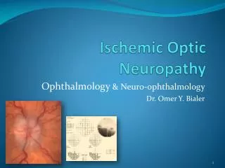
Ischemic Optic Neuropathy. Ophthalmology & Neuro-ophthalmology Dr. Omer Y. Bialer. Disclosure. No conflict of interests I have nothing to disclose ION = I schemic O ptic Neuropathy. Presentation’s outline. Introduction Terminology and Nosology

E N D
Ischemic Optic Neuropathy Ophthalmology & Neuro-ophthalmology Dr. Omer Y. Bialer
Disclosure • No conflict of interests • I have nothing to disclose ION = Ischemic Optic Neuropathy
Presentation’s outline • Introduction • Terminology and Nosology • Nonarteritic anterior ischemic optic neuropathy • Arteritic ION • Perioperative ION • Radiation optic neuropathy • “Take home massage” summary
Introduction • ION is the most common acute optic neuropathy > age 50 • 2nd most common optic neuropathy after glaucoma • Relatively common neuro-ophthalmological disorder • Visual loss is often severe • No effective treatment or prevention
Introduction • ION is due to: • poor blood flow to the optic nerve • Acute occlusion of the feeding arteries Ophthalmic artery Short posterior ciliary arteries
Terminology & Nosology ION Nonarteritic ION (cardiovascular risk factors) Arteritic ION (vasculitis) NonarteriticAnterior ION (NAION) with swollen optic disc NonarteriticPosterior ION (NA-PION) with normal optic disc Arteritic Anterior ION (AAION) with swollen optic disc Arteritic Posterior ION (APION) with normal optic disc
Terminology & Nosology ION Nonarteritic ION (cardiovascular risk factors) Arteritic ION (vasculitis) NonarteriticAnterior ION (NAION) with swollen optic disc NonarteriticPosterior ION (NA-PION) with normal optic disc Arteritic Anterior ION (AAION) with swollen optic disc Arteritic Posterior ION (APION) with normal optic disc GCA Other vasculitides Idiopathic ION Perioperative ION Radiation optic neuropathy
NAION (Nonarteritic Anterior Ischemic Optic Neuropathy)
NAION is the most common ION • ~ 90% of ION • Incidence: 1 / 10,000 / year (> 50 y.o) 0.5/ 100,000 / year (overall) • Mean age at onset 57-65 • Presentation: acute painless monocular visual field loss ± visual acuity loss
The most important risk factor is a crowded optic disc • “disc at risk” = small optic disc + minimal cup crowded normal glaucoma
More risk factorsfor NAION • Hypertension (50%) • Diabetes mellitus (25%) • Obstructive sleep apnea (55%) • Hyperlipidemia • Ischemic heart disease • Obesity • Tobacco use • High intraocular pressure
Several meds are associated with NAION • Erectile dysfunction drugs • Amiodarone • Vasoconstrictors • Cocaine (e.g. Viagra, Cialis) (e.g. nasal decongestants)
The pathogenesis of NAION differs from IHD or CVA Edema of optic disc Cardiovascular risk factors decrease in blood flow Compression of axons and blood vessels Crowded optic disc Blockage of axonal flow Necrosis and demyelination of nerve fibers
Eye Exam • visual acuity & color vision can be normal • A relative afferent pupillary defect • Normal anterior segment • Optic disc edema • Crowded optic disc (fellow eye) Peripapillary hemorrhages Obscured borders Nerve fiber layer edema
The most common visual field defect is a superior or inferior scotoma Combined superior & inferior defect Inferior altitudinal defect Superior arcuate defect
NAION is a clinical diagnosis • Elderly patient +/- cardiovascular risk factors • Acute painless optic neuropathy + disc edema + crowded optic disc in fellow eye • Rule out arteritic AION • Do Humphrey visual fields • Imaging is not in indicated • Frequent follow-up
There is no proven treatment for NAION • IONDT = ION decompression trial • A multicenter randomized controlled clinical trial • no efficacy for optic nerve fenestration • Intravitreal steroids (triamcinolone acetate) • Intravenous noradrenaline • Warfarin • TPA • Levodopa + carbidopa
There is no proven treatment for NAION • Oral prednisone 40-60mg daily – may hasten resolution of disc edema • Some evidence for anti-VEGF intravitrealinjections
Prophylaxis • Control of cardio-vascular risk factors • Aspirin 100 mg daily – limited evidence for second eye prophylaxis
Disc edema resolves in 1 month cup Optic atrophy with cupping Optic atrophy
Significant improvement is rare • ~40% experience partial improvement • Improvement may take up to 6 months • 15% risk for fellow eye involvement in 2 years • < 5 % recurrent AION (the same eye) • A significant visual field defect persists
Arteritic ION And Giant Cell Arteritis (GCA)
>50% of Arteritic ION are d/t Giant Cell Arteritis • Other etiologies include: • Systemic Lupus Erythematosus • Wegener’s granulomatosis • Behcet’s disease • Churg Strauss • PolyarteritisNodosa
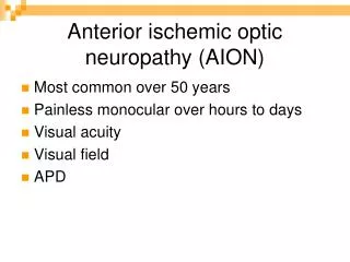
GCA* - key facts • Large vessel vasculitis • Predilection for the aortic arch • Incidence 20 / 100,000 / year (> age 50) • 20% of GCA patients experience severe visual loss • AION is the most common ophthalmic manifestation of GCA • A-AION is an ophthalmic emergency ! * GCA = Giant Cell Arteritis (Temporal arteritis)
Arteritic ION presents like any ION, but . . . • 75% have typical systemic symptoms • 30% have preceding transient visual loss • 54% have visual acuity of count-fingers No light perception • >50% second eye ION within hours -weeks (“amaurosisfugax”) (vs 26% in NAION)
There are specific funduscopic findings The involved swollen optic disc is acutely pale NAION
There are specific funduscopic findings Ischemic retina Cherry red spot Branch Retinal Artery Occlusion Central Retinal Artery Occlusion
There are specific funduscopic findings Lack of choroidal perfusion normal choroid Choroidalhypoperfusion indicates multifocal ischemia on Fluorescein angiography
The workup of suspected Arteritic ION GCA Symptoms / signs ? Do blood tests but yes no ESR, CRP, Hb, PLT, Fibrinogen IV Solomedrol Prednisone + aspirin until biopsy results Iv Solomedrol Prednisone + aspirin NAION high normal Urgent TAB* TAB* in 1 w * TAB = Temporal Artery Biopsy
“Ophthalmic GCA” should be treated with IV steroids • Few studies evaluated treatment protocols • Studies in ophthalmology differ from rheumatology • We recommend: • IV methylprednisolone 1000mg/d for 3 days • followed by a very slow taper of oral prednisone • Aspirin 100mg daily • Rheumatology consultation & follow-up
Perioperative ION (post operative AION and PION)
ION is a rare surgical complication • ION is an uncommon but devastating complication after various types of surgeries • Intraocular surgeries • Intraocular injections • Non-ocular surgeries • ION may also occur after: • renal dialysis • cardiac catheterization d/t Elevated intraocular pressure
ION may complicate non-ocular surgeries • The 2 most “classic” are : • CABG • Spinal surgery • Commonly bilateral • There is often profound visual loss • Visual loss may be immediate or delayed (days) (mostly AION, 0.06%) (mostly PION, 0.2%)
The differential diagnosis of post-operative visual loss includes • Ischemic optic neuropathy • Retinal artery occlusion • Angle closure glaucoma Cherry red spot Hazy cornea Unresponsive mid-dilated pupil Red “angry” eye
The differential diagnosis of post-operative visual loss includes • Cortical blindness • Corneal erosion Bilateral occipital stroke Epithelial irregularity
There is no prospective / controlled data regarding perioperative ION • Risk factors: • Obesity • Male gender • Prolonged surgical time • Surgery in the prone position • Large fluid shifts / severe blood loss
There is no effective treatment • Prognosis is poor – significant improvement in minority of patients • Should correct anemia, saturation & hypotension to improve perfusion • No evidence for efficacy of : • Aspirin • Anti - coagulants • Thrombolytics • Anti-glaucoma drops
RON (Radiation Optic Neuropathy)
RON is a late complication • Prevalence ~ 0.5% • Mean interval 18 months • The optic nerves must be in the radiation field • (range: 3 months – 9 years)
The risk factors are: • Radiation dosage • Age • Diabetes mellitus • Presence of compressive optic neuropathy • Concomitant chemotherapy • Previous radiotherapy • Multiple sclerosis • (>total 50 Gy or single dose > 10 Gy)
RON mostly presents as PION • May be monocular or binocular • 45% have visual acuity of no light perception • Diagnosis is one of exclusion: • Suspected Optic neuropathy • PMH of radiotherapy • No other obvious explanation • Optic nerve enhancement on MRI
Isolated enhancement on MRI optic nerve enhancement T1W with fat suppression + gadolinium
There are few treatment options • Oral corticosteroids (prednisone 1mg/kg) • Anticoagulants (heparin) • Aspirin • Hyperbaric oxygen (30-60min/day x 14-30 days) • Intravenous Bevacizumab (2-4 cycles every 2 weeks)
Suspected RON ? Onset < 48-72 hours ? yes no VEP Look for other etiologies Brain+orbits MRI with gadolinium normal abnormal Hyperbaric oxygen yes PO prednisone Enhancement ? Consider IV Bevacizumab Other optic neuropathy
Prognosis of RON is poor • Spontaneous recovery is rare • Treatment is mostly ineffective • 85% visual acuity ≤ 20/200 • Optic atrophy appear in 6-8 weeks • Enhancement on MRI resolves after several months
Conclusions (the “take home massage”)
ION is an ophthalmic emergency • Patients with GCA+ION are in danger of catastrophic, irreversible, bilateral blindness that may be prevented by prompt treatment with corticosteroids • Any patient > 50 presenting with ION an immediate workup to rule out GCA
ION is not “another type of CVA” • Although considered a “stroke of the optic nerve” and shares many risk factors with cerebrovascular disease, It cannot be directly compared to cerebral infarction, and therefore the evaluation should not be similar to that of cerebral infarction.
There is no effective treatment for ION • there are no class I studies showing benefit from any medical or surgical treatments TPA Steroids Anti VEGF Heparin Aspirin Levodopa Erythropoietin Decompression surgery Noradrenalin Hyperbaric oxygen
Limited efficacy for prophylaxis • Aspirin 100mg daily • Control of cardiovascular risk factors • suspect GCA !!! • Avoid prolonged surgical time and dramatic shifts in body perfusion during surgrey • Consider routine serial brain MRIs after brain radiotherapy to detect RON early