Download
1 / 28
290 likes | 1.99k Views
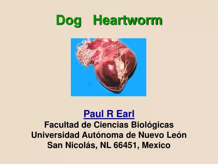
Dog Heartworm Paul R Earl Facultad de Ciencias Biológicas Universidad Autónoma de Nuevo León San Nicolás, NL 66451, Mexico.

E N D
Dog HeartwormPaul R EarlFacultad de Ciencias BiológicasUniversidad Autónoma de Nuevo LeónSan Nicolás, NL 66451, Mexico
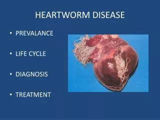
Dog heartworm disease (dirofilariasis) of dogs and other canids like foxes also infects cats but to a much lesser extent. It is almost worldwide and often common in humid areas that support mosquitoes as in the entire Mississippi valley. Rarely as in Florida, it can infect man causing pulmonary infarction with granuloma formation. Before the 1960s, Dirofilaria immitis (Leidy, 1856) Railliet & Henry, 1911 was best known along the northern Gulf of Mexico, Florida and along the entire east coast of the US. D. immitis is still colonizing northern Europe and England, perhaps in search for suitable local mosquito vectors. Note that its larvas = microfilarias = mfs have a marked nocturnal periodicity in the peripheral blood.
The taxonomic groups are Genus & Species Dirofilaria immitis of Family Onchocercidae of Order Spirurida of Subclass Spiruria of Class Secernentea. Mosquito-borne D. repens inhabits the skin of dogs. Transmitted by fleas, Dipetalonema (= Acanthocheilonema) recondita also inhabits the dog's subcutaneous tissues. However, some of this kind have biting midges Culicoides spp. as vectors. Perhaps Dirofilaria, Dipetalonema and Acanthocheilonema are synonyms. The proper systematics for Dirofilaria awaits nucleotide sequencing using the polymerase chain reaction (PCR). The complete mitochondrion squence is already known for D. immitis.
By clogging the main blood vessels, the blood supply to other organs of the body is reduced, particularly the lungs, liver and kidneys.Of course, much more work for the heart results. Therefore, tachycardia (rapid pulse) and dysnea (shortness of breath) are typical. Blockage of major blood vessels can cause the animal to collapse suddenly and die within a few days. Sudden destruction of large numbers of mfs occasionally causes severe shock-like symptoms that may kill the dog. Most dogs can be treated successfully if heartworm disease is detected early by blood tests. Other diagnostic tests include radiography and electro/echo-cardiography.
Dipetalonema recondita.The adult worms measure 13 mm (male) to over 25 mm (female). Fleas as Ctenocephalides felis and lice as Heterodoxus spiniger are the intermediate hosts. The flea ingests mfs when feeding on an infected dog. The mfs develops into infective larvas in 7-14 days. When the flea again feeds on a dog, the larvas are injected into the skin. The female worm lays mfs which find their way into the bloodstream. The prepatent period is about 60-65 days. As the host is asymptomatic, there is no real treatment!
Heartworm disease in dogs & cats and Angiostrongylus infectionEtiology. The nematode Dirofilaria immitis infects mainly dogs but also cats, ferrets and some other animals, including--rarely--man. The mfs in the blood can be about 315 long by 6-7 microns in diameter.Distribution. It has a worldwide distribution, and the disease is most common in dogs in humid areas like Florida. The distribution follows that of the mosquito vector. See the map above ! Life Cycle. The worms mature in dogs in 6-7 months, adults living in the chambers of the heart and the pulmonary arteries. Adults mate and large numbers of mfs are discharged into the blood stream. Mfs can survive for 1 - 3 years. The mfs are ingested by a mosquito.
Mosquito Vectors.Many mosquitoes worldwide have been identified as capable of sustaining the development of dog heartworm mfs to the infective stage. Examples are Aedes aegypti, Ae. albopictus, Ae. canadensis, Ae. cantator, Ae. excrucians, Ae. sollicitans, Ae. sticticus, Ae. stimulans, Ae. taeniorhynchus, Ae. vexans, Anopheles bradleyi, A. punctipennis, An. quadrimaculatus, Culex nigripalpus, Cx. quinquefasciatus, Cx. salinarius and Psorophora ferox have been identified as natural hosts of D. immitis in the US east of the Mississippi River. Among these, 11 species are found in abundance in Florida.
Clinical signs and pathologyDogs with low numbers of adult wormsmay be asymptomatic and also produce false negatives. Chronic disease may develop in more infected dogs. Coughing, reluctance to exercise, weight loss, weakness and dypsnea are clinical signs. With heavy infections, there may be circulatory distress-dypsnea due to reduction of blood flow and pulmonary hypertension. Typically, signs include coughing, labored breathing, weakness and tiring with exercise. Since the signs vary, the disease may be well advanced before the dog begins to show problems. In advanced stages, the heart, lungs, liver and kidneys may be severely damaged. Eventually, heart failure can occur involved with overwork by the heart.
DiagnosisHeartworm disease should be suspected in dogs a year or more of age with some of the signs mentioned above in endemic regions. This disease is usually detected by finding mfs in a blood sample by either 1/ looking for mfs with a microscope at 20-100 x or 2/ using one of several serological diagnostic tests which can detect of heartworm antibodies even when mfs cannot be found with a microscope. A drop of blood is smeared on a slide, dried, fixed with 95 % ethanol and stained with Giemsa or Wright's, as in standard hospital procedure. Very simple. However, unstained fresh blood or blood with anticoagulant can reveal mfs by their writhing movement at very low power.
D. recondita mfs stain rather solidly with acid phosphatase, whereas D. immitis mfs produce a few spots.
The respective mfs have these sizes: D. recondita 264 x 4.4, D. immitis 300 x 5.0 and D. repens 325 x 7.2 microns. Length difference 12 % between D. recondita and D. immits, and D. recondita and D. repens of 19 % are sufficient to differentiate these mfs, whereas the other differences are not.The differential diagnosis of D. Immitis & D. recondita has been worked out using PCR with primers for the internal transcribed spacer region 2 (Vet Parasit 106: 243-252, 2002).In a fresh thin blood smear the mfs of D. immitis undulate in one place while those of D. recondita move across the field of the microscope.
Knott's technique: A/ 1.0 ml freshly drawn blood added to 10.0 ml of 2 % formalin, B/ Shake to help lyse the RBC, C/ Centrifuge 5 min at 1000-1500 rpm, D/ Decant, E/ Stir residue with wooden applicator to free the mfs from the walls of the tube and F/ to sediment on a slide, add an equal volume of 1/1000 (or less) methylene blue or acridine orange; after a few minutes mount a drop under a coverslip.D. recondita has a characteristic button-hook tail.
The acid phosphatase test is the most accurate method for differentiating the mfs of D. immitis, D. repens and D. recondita. Mfs from serum, after clotting the blood, are air-dried on a slide. The slides are then incubated for 1 hour in a substrate of Michaelis vernal acetate buffer (pH 10.0), naphthal AS-TR phosphate, pararosaniline and 4.0 % sodium nitrate. After rinsing in distilled water, it is counterstained with methyl green, rinsed successively in 95% ethanol and absolute ethanol, and mounted in Permount.
Treatment. Thiacetarsamide (Caparsolate) and melarsamine (Immiticide) are arsenical type compounds that consistently kill the adults. Mfs can be killed with ivermectin. This drug is supreme. Many other drugs are very successful, the problem being dead worms & mfs whose presence in the dog's lungs is downright dangerous.
Ivermectin: (Heart Guard, Iverhart, Stromectol, Mectizan)--Macrocyclic lactone derivative of avermectin (22, 23-dihydro-avermectin). Binds selectively with glutamate-gated chloride ion channels in invertebrate nerve and muscle cells, causing cell death. Half-life is 16 h; metabolized in liver. Exerts its antiparasitic action by acting as a potent agonist at GABA receptors and potentiating the inhibitory signals sent to motor neurons, which paralyses the parasite. As GABA is confined to the central nervous system in humans and ivermectin does not cross the mammalian blood-brain barrier, the drug has no paralytic action in humans. Adult Dose: 150-200 mcg/kg administered orally.
Management can consist of the following: 1/ diagnostic evaluations (before therapy) to determine subclinical disease, especially of the liver and kidneys; 2/ adulticidal therapy to eliminate mature worms; 3/ a rest period of 4-6 weeks to allow the dog to recover from the lung injury associated with worm death; 4/ mf therapy; 5/ a test for mfs to determine success of mf therapy; 6/ an antigen test to determine success of adulticidal therapy and 7/ preventive medication. The death of the worms is associated with parenchymal lung damage. Shock from dead mfs is another hazard. Limitation of exercise is critical after adulticidal therapy.
Complications of worm death often include impaired pulmonary function and vessel damage, which may initiate disseminated intravascular coagulation (DIC). A platelet count <100,000/ml is not uncommon, but an activated clotting time should be performed to determine if DIC is present. If so, the prognosis is poor. Antigen assays 12-16 weeks after successful adulticidal therapy should be negative. However, low worm burdens and immature worms can still be present with a negative antigen test. Positive antigen tests should be rechecked in 1 month before initiating a 2nd adulticidal regimen. This is deluxe treatment.
Surgery avoids embolism and other ills. Rather than destroy heartworms with arsenicals and other drugs, it is sometimes better to physically remove them from the pulmonary arteries. This treatment allows for removal of most of the worms without risking complications due to thromboembolism. Then, dogs with severe cor pulmonale or caval syndrome will benefit greatly. As worms are removed from the pulmonery artery with forceps, more will come up from the heart. Close your incision in the artery with a clamp to minimize escaping blood. Remove it. Then fish out more worms in the artery. However, there are several ways this operation can be conducted, using alligator flexible forceps and so on.
Control. Mosquito control depends upon public health (PH) measures per district and per season. PH protects the public by indoor & outdoor spraying, and by mosquito larva control, not by killing the parasite. Repelents are hardly common. Deet repelent is good for you and bad. for your dog.Is heartworm disease effectively prevented by once monthly medication beginning at about 2 months of age? Yes. In tropical regions, medication is administered throughout the year; in temperate areas prophylaxis is begun about one month before the mosquito season and concluded about a month after.
Flea collars as preventing heartworm disease have never been studied !They contain organophosphates and protect against leishmaniasis and all other insect borne diseases. They kill fleas, mosquitoes, triatomid bugs, ticks, etc. Flea collar insecticides have a VERY SLOW evaporation that is PERFECT for ridding the dog's kennel and your home from these pests. Vaporizers that plug into an electric outlet. are also PERFECT for clearing houses of vectors and pests like cockroaches. Still, even perfect has limits !
Eradication. This requires a level of cooperation by no means evinced! All dogs & cats in animal shelters must be dosed at least once as well as all licenced animals to clear mfs. Organophosphates go after vectors, whereas a great variety of drugs attack the parasites. Should the financing be raised in some area like highly infected Florida to successfully eradicate heartworm? Would infected endemic wild animals bring back dirofilariasis ?Veterinary epidemiology does not concentrate on vector control and surveillance, possibly because these underinvestigated fields are not financed. For example, no vaccine development is based on low profits. The motivation to eradicate dirofilariasis has not yet arisen.
Public Health Significance Infection results from mosquito bites. The worms lodge in the pulmonary artery causing vascular occlusion, coagulation, necrosis and fibrosis. Symptoms include chest pain, coughing and in some cases hemoptysis. In some patients radiographs have disclosed an asymptomatic, fibrotic nodule 1-3 cm in diameter.
Heartworm disease of catsCats are susceptible yet are poorer hosts than dogs. The most prominent clinical signs include coughing, dyspnea, vomiting, lethargy and anorexia. Acute collapse and death can occur. As less than 20% of infected cats have mfs in the blood, diagnosis is best confirmed by either X-rays, echocardiograhy or serologic tests.Etiology. Heartworm disease caused by Dirofilaria immitis is a significant disease of cats. It is found worldwide and in many states in the US where the canine disease occurs. Prevalence rates in cats are estimated to be about 10 % of that of local dogs. The average worm burden is 1-5. The mean age of affected cats is 3-6 years.
Angiostrongylus infectionEtiology. This parasite is the nematode Angiostrongylus vasorum, up to 2.5 cm long.Distribution. With the exception of North and South America, it is found worldwide. Because of the infection’s frequency in France, it is called the "French heartworm." Dogs are the definitive host and adults are found in pulmonary vessels. It has been reported in dogs imported into the US.