Download
1 / 83
1.53k likes | 5.4k Views
Hematological disorders in pregnancy. Guide- Dr. Neeta Singh CO-guide- Dr. Sujata Rawat Candidate- Dr. Prerna. Headings. Disorders of RBC’S – Anemia, Hemoglobinopathies & polycythemia Disorders of WBC’s Disorders of Platlets Coagulation disorders – Inherited/ Aquired

E N D
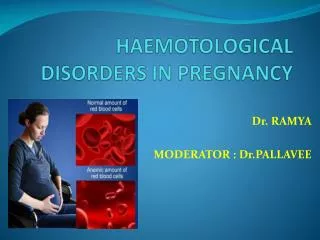
Hematological disorders in pregnancy Guide- Dr. Neeta Singh CO-guide- Dr. SujataRawat Candidate- Dr. Prerna
Headings • Disorders of RBC’S – Anemia, Hemoglobinopathies & polycythemia • Disorders of WBC’s • Disorders of Platlets • Coagulation disorders – Inherited/Aquired • Hematological malignancies
Anemia ANEMIAS OF DECREASED RBC PRODUCTION ANEMIA DUE TO RBC DESTRUTION • DECREASED Hb SYNTHESIS- MICROCYTIC • IRON DEFICIENCY • THALASSEMIA • SIDEROBLASTIC ANEMIA • DECREASED DNA SYNTHESIS- Megaloblastic anemia • STEM CELL FAILURE – Aplastic anemia • ANEMIA OF CHRONIC DISEASE • Hemolytic anemia • Autoimmune • Hemoglobinopathies
Hemolytic anemia • Premature destruction of RBCs - inherited defects/acquired intravascular abnormalities. • Hemolysis -Intravascular or extravascular • General features of hemolytic anemia
Intravascular destruction of RBCs schistocytes Causes- mechanical trauma, complement fixation, toxic damage to the RBC. Decreased serum haptoglobulin, hemoglobinemia hemoglobinuria, hemosiderinuria Iron loss
Extravascular destruction of RBCs Causes -bound immunoglobulin, or physical abnormalities restricting RBC deformability that prevent egress from the spleen. Iron overload leading to secondaryhemochromatosis-damage to liver& heart
Clinical features Due to anemia • Weakness, exhaustion & lassitude, indigestion, loss of appetite • Palpitations, giddiness, dyspnoea • Pallor, hyperdynamic circulation, flow murmur Due to hemolysis • Icterus, splenomegaly in extravascularhemolysis, gall stone disease Lab investigations in hemolytic anemias • Complete hemogram with reticulocyte count PBS – anemia with reticulocytosis & fragmented RBC on PBS • Decreased serum haptoglobulin. • LFT/KFT • DCT • USG abdomen- Hepatosplenomegaly • HPLC, Osmotic fragility test
Membrane disorders-Hereditary spherocytosis • MC -northern European ancestry • AD, chr8p • Defect- abnormality of ankyrin. • Decreased surface area/vol-Spherocytes - less flexible – extravascularhemolysis-splenomegaly. • Only condition with increased MCHC. • Increased osmotic fragility (Pink test) • TT- No tt aimed at cause. • Splenectomy – Obligatory . • In pregnancy (rare)- fetal loss in 1st trimester, aplastic or hemolytic crisis, increased folic acid requirement due to chronic hemolysis • Splenectomy – 2nd trimester • Affected Fetus – neonatal jaundice, need for exchange transfusion • PND- by CVS, amniocentesis Normocytes, Spherocytes.
ENZYME DISORDERS-G-6-PD deficiency • MC enzyme deficiency . • X linked recessive • Mediterranean, West African, Mid-East, and Southeast Asian populations • Interaction between extracorpuscular & intracorpucular cause. • Heterozygotes - resistant to P falciparum. • Oxidative stress- Increased methemoglobin, aggregates of denatured hemoglobn to form heinz bodies, membrane injury • Screening – NADPH mediated dye decoloration • Diagnosis – spectophotometric assay of NADPH production, G6PD enzyme assays
Contd……. In pregnancy: • Spontaneous abortion, still birth & low birth wt babies with neonatal jaundice • Affected fetus – non immune hydrops if mother ingests oxidant drugs crossing the placenta • PND- CVS or FBS • Avoid agents causing hemolysis • Acute hemolytic episode – adequate hydration, maintain urine output, BT if needed • spherocytes, schistocytes, bite cells & blister*
Pyruvatekinase deficiency • Autosomalrecessive • Reduced ATP formation causes RBC membrane rigidity. • PBS- polychromasia, anisocytosis, poikilocytosis with burr cells & acanthocytes • Symptoms -usually mild (right shift of the 02-dissociation curve). • Homozygote-severe anemia & usually discovered in childhood. Splenomegaly, cholelithiasis and jaundice . • In pregnancy – well tolerated, supportive management during crisis & BT if needed • Splenectomy – 2nd trimester • Fetus– nonimmunehydrops. FBS for diagnosis & IUT if needed
MECHANICAL TRAUMA • RBCs striking against abnormal surfaces (aortic stenosis,atherosclerosis) or artificial surfaces (prosthetic heart valves; arterial grafts). • Microangiopathic hemolytic anemia - RBCs torn apart on fibrin strands strung across small vessels or on damaged endothelial surfaces of small vessels. • Accompanies DIC, malignant hypertension, HUS, TTP, pre-eclampsia, and some vascular neoplasms.
HEMOGLOBINOPATHIES • Abnormalities due to alteration in structure, function or production of hemoglobin • inherited disorders- autosomal dominant (unstable hemoglobins) and autosomal recessive (Hgb S). • The most common are thalassemia and sickle cell disease/trait. • Minor disorders • Sickle cell trait- Hb AS • Hb SE disease • Hb SD disease • Hb S Memphis • Major disorders • Sickle cell anemia – Hb SS • Hb SC disease • Hb S ß thal
Sickle cell Disease • Qualitative disorder • Point mutation in the ß-chain at codon 6 encoding of a valine instead of normal glutamin. • Hb S- poorly soluble in low oxygen tension, polymerizes into fibrilary structures/ tactoids-- causing them to become rigid and sickled. • M.C inherited hematological disease worldwide • Most prevalent in African descent(1 in 625). ACOG technical bullein, no. 185, Oct 1993 The term sickle-cell disease is preferred because it is more comprehensive than sickle-cell anaemia.
Autosomal recessive Homozygous/Heterozygous(coinheritance with other abnormal hemoglobin ; mostcommonlyHbSC or b thalassemia) Diag-chances (for each pregnancy)of two carrier parents having a child with a sickle cell or thalassaemia disorder.
If the mother is anemic & the father is healthy carrier 50% of the off springs are carriers and 50% is anaemic
PATHOPHYSIOLOGY • Hemolysis • Vaso-occlusion-because of- a)sickled cells are less deformable& more fragile & also have increased tendency for cellular dehydration b) Increased adhesion of red cells to vascular endothelium(increased expression of adhesion molecules, upregulation of thrombotic pathways, proinflammatory state) • Life span of sickle cells – 17 d Initially, oxygenation restores normal shape. With repeated cycles of agglutination & polymerisation, sickling becomes irreversible
Diagnosis • HIGH PRESSURE LIQUID CHROMATOGRAPHY • Isoelectricfocussing • Cellulose acetate electrophoresis at alkaline pH • Capillary electrophoresis • Sickle cell solubility test- Widely used screening method. • Relies on the relative insolubility of Hgb S in concentrated phosphate buffers compared to Hgb A and other Hgb variants. Hgb S precipitates causing a cloudy solution.
Sickle cell trait • Hgb SA- 25 - 45% of the hemoglobin is Hgb S; remainder being Hgb A, Hgb F & HgbA2. • No anemia and normal RBC morphology is the rule. • Two rare complications-hematuria and splenic infarction. • No risk from anesthesia, surgery, pregnancy, or strenuous physical activity. • Normal growth & development, normal life spans • Increased incidence of pre-eclampsia in pregnancy
Preconceptional care General advice & care - • At least annual review at specialist clinic - • BP measurement, • KFT testing, • ophthalmological checkup, • screening for red cell antibodies & iron overload*, • cardilogy review for pulmonary hypertension( echo not done in last year) • Specific issues in women trying to conceive- counselling about • Risk of worsening anemia, increased infections(especially UTI), pain, IUGR, PTL, Pre-eclampsia , caesarean section & perinatal mortality. • Role of dehydration(Early detection & treatment of nausea &vomiting), cold, hypoxia, overexertion, &stress in frequency of sickle cell crisis
ANTENATAL SCREENING Pregnancy Offer screening Blood sent to laboratory for haemoglobinopathy Screen Negative Result Information: No further action Positive results Information & counseling-Offer partner screening Partner screening Blood sent to laboratory for haemoglobinopathy Screen Negative Result Information: No further action Positive results: At risk couple Information & counseling-Offer prenatal diagnosis Prenatal diagnosis Fetal blood Sampling/ Chorionic Villus sampling Unaffected Fetus Information- No further action Affected fetus-Information &counseling Continue with Pregnancy Parents Make- Informed Choice Termination of Pregnancy
Sickle cell disease contd…. • Medications-Daily penicillin prophylaxis (250 mg BD) • Folic acid 5mg once daily throughout pregnancy • Hydroxycarbamide should be stopped 3 months prior to conception( termination is not indicated based on exposure to hydroxycarbamide alone). • ACE inhibitors & Angiotensin 2 receptor blocker , iron chelating agents should be stopped • NSAID’s are not recommended <12weeks& >28 weeks ; should only be taken after medical advice in 2nd trimester. • Vaccinations ( preconceptional)*-H.influenza type b, conjugated meningococcal C vaccine, Hepatitis B , Influenza & swine flu vaccine annually, pneumococcal vaccine every 5 years. Management of sickle cell Disease in pregnancy. RCOG2011
Sickle cell disease contd…. • Indications of urgent transfusion therapy- 1)Acute anemia - top up transfusion, • Hb <6 g/dl or • a fall of over 2g/ dl or • symptomatic patients. 2)Acute chest syndrome& acute stroke – exchange transfusion Role of prophylactic transfusion in pregnancy- • Insufficient evidence to draw the conclusion about role of prophylactic blood transfusions in pregnancy. Mahomed K et al. prophylactic versus selective blood transfusion for sickle cell anemia.2006. The cochrane library, issue 2. • Indicated for women who are on a long term transfusion regime prior to pregnancy.*
Antenatal care • Multidisciplinary team • Pregnancy– exacerbations of disease manifestations • increased metabolic demands, • hypercoagulablestate, • increased vascular stasis – • Vaso-occlusive crisis – common in later half of pregnancy • Pyelonephritis – altered immune system added to renal changes of pregnancy • Symptomatic cholelithiasis – chronic hemolysis, progesterone induced changes in GIT • Susceptible to infections, pre-eclampsia,thromboembolism • IUGR, preterm labour, abruption, • SCREENING – selective (low preevalance area) versus universal(high prevalance area) mainly to diagnose minor forms. If positive, screen partner, genetic counselling, PND
Antenatal management • Early booking • ANC Visits monthly upto24weeks, 2weekly until 36weeks& weekly thereafter. • Low dose aspirin (75mg daily )from early pregnancy(12weeks) till 28weeks. • Routine thromboprophylaxis only if they have additional risk factors, but should receive LMW heparin during antenatal hospital admission. RCOG 2009. Reducing the risk of thrombosis in pregnancy & puerperium. Green top guideline 37. Role of iron suplementation: • Iron supplementation is withheld unless there is e/o iron deficiency. Akien’ova YA et al. Ferritin & serum iron levels in adult patients with sickle cell anemia in Ibadum, Nigeria. Afr J Med Sci1997;26.
Contd………. • Routine iron supplementation entails a negligible theoretical risk of iron overload for a substantial benefit. Streetly A et al, BMJ 2000; 320. • BP & Urinalysis at each visit. • Serial USG for GP & AFI from 24 weeks; every 4 weekly& more frequently if there is evidence of poor growth. • Monthly assessment of hct, reti count, urine c/s • Fetal monitoring – DFMC, weekly NST & BPP • Maintain oral hydration, diagnose & treat infections early • Mode of delivery- In the absence of obstetric indications allow spontaneous labour at term • Role of cytotoxic agents to HbF & HbA– 5-azacytidine, hydroxyurea – investigational in pregnancy. `
Management of acute painful episodes during pregnancy • Most frequent complications, incidence- 27%-50%. • Mild pain –rest, fluids & simple analgesia(paracetamol& week opoids) • Severe pain- low threshold for admitting to hospital. • Assess for other complications precipitating factors(Dehydration) • Ix-spo2, urinalysis, full blood count, reticulocyte count, KFT, Urine c/s, blood c/s, chest x-ray. • Tt- strong opoids- morphine/ diamorphine(oral/parenteral) are the first line agents. • Give adjuvant non-opoid analgesia: PCM, NSAIDS(12-28weeks) • Monitor for pain score, sedation score, & oxygen saturation using a modified obstetric early warning chart(MEOWS), RR every 20-30minutes until pain is controlled & signs are stable, then monitor every 2 hour or hourly if receiving parenteral opiates. • Give rescue dose of analgesia if required. Rees DC et al. Guidelines for the management of acute painful crisis in sickle cell disease. BJH. 2003;120.
Contd… • If RR<10/min, omit maintenance analgesia; consider naloxone • Oral/ iv fluids – 60 mg/kg/24 hours.(precaution – PET) • Maintain I/o chart • Antibiotics & Thromboprophylaxis should be used. • Consider reducing analgesia after 2-3days & replacing injections with equivalent dose of oral analgesia. • Discharge when -pain is controlled/ improving without analgesia or on acceptable doses of oral analgesia. Rees DC et al. Guidelines for the management of acute painful crisis in sickle cell disease. Br J Hematol 2003;120.
Intrapartum management • Timing of delivery- 38-40 weeks. • Mode of delivery- vaginal. • Adequate hydration • Pulse oxymetry should be used throughout labour • Supplemental oxygen therapy used if necessary to maintain spo2 >94%. • Antibiotic therapy should be used if there is evidence of, or high clinical suspicion of infection. • Continuous fetal monitoring • Epidural analgesia • Regional anaesthesia preferred for caesarean section. • Hourly vital signs- low threshold to start broad spectrum antibiotics Management of sickle cell Disease in pregnancy. RCOG2011
Post partum management • Risk of thrombo-embolism, painful crisis • Early ambulation, hydration,painreleif (NSAIDS/pcm/ opoids) • Prophylactic sucutaneousLMW heparin for 7 days after vaginal delivery & 6 weeks following a caesarean section. • Aggressive treatment of suspected infection • Cord blood – HPLC • Encourage breast feeding • Antithrombotic stocking • Baby affected- prophylactic penicillin from 3 months of age- ↓ incidence of pneumonia. • Contraception- progesterones are effective & safe contraceptive . First line- PIC, MIRENA, Implanon, pop, barrier method. Second line- COC, Cu- IUD, Vaginal ring , Combined patch.
Imbalance of globin chains available for hemoglobin dimer construction. ß thalassemia- defective synthesis of the ß chain. A thalassemia, defective synthesis of the a chain (quantitative). Globin chain (a, b, d, e, g & z) structural genes are located on chromosome 16 (a;z) and chromosome 11 (b;d; e;g). THALASSEMIAS
Geographic distribution • ß -thalassemia is common in the Mediterranean region, Africa, Asia, the South Pacific, and India. • a -thalassemiamore common in Southeast Asia. • Prevalance- 16% in southern European , 10% in Thiland , 3-8% in Indian , Pakistani & Bangladeshi population Leung TN et al. Thalassemia screening in pregnancy.CurrOpinObstetGynecol 2005; 17.
point mutations or a partial deletions of chromosome 11 cause defective synthesis of the ß chain.( >100 mutations) Normally- a and b globin chains are roughly equal amounts. When ß-globin chains are in short supply or absent, the excess a-chains combine with other available ß-family globin chains ( d or g) to form increased amounts of HgbA2 (a2d2) & HgbF(a2g2). HgbBarts( g4) or tetramers of excess gamma chains may form. ß-Thalassemia
The clinical severity depends on the degree to which production of the ß-chain is inadequate. • ß-thalassemiamajor-no ß chains (ßo) or very little is made (ß+). • ß-thalassemia minor-ß+ chains are made in mildly reduced amounts. • ß+thalassemiaintermediaß+ chains are made in amounts intermediate to the major and minor forms. Signifcance of ß-gene Mutation • type 1 ß+ - about 10%of normal ß chain production • type 2 ß+ -about 50%of normal ß chain production • type 3 ß+ - >50%of normal ß chain production
ß-Thalassemia major • No ß chains (homozygous for ßo, Cooley's anemia), or very little ß chain (homozygous for ß+). • Hgb electrophoresis-↑HbF,↑HbA2, variable amounts of Hb A. • PBS - severe anisocytosis& poikilocytosis, targets, elliptocytes, teardrops • Asymptomatic till 6 months of life** • C/F- severe, transfusion dependent anemia. Nearly all have hepatosplenomegaly. • Expansion of the marrow by erythroid hyperplasia - enlargement of bones. • Iron overload, secondary to transfusion dependency, results in damage to the heart, liver and endocrine organs. • Short life span, most dying before adulthood.
ß-Thalassemia minor • ß-thalassemiatrait/ minor- Heterozygous- mildly reduced production of ß+ chains & thus, a mild excess of a globinchains which denature, causing damage to young red cells in the marrow (ineffective erythropoiesis) or decreased survival in the peripheral blood. • ß-thalintermediamb homozygous for type 2 ß+ and type 3 ß+. • Mild anemia • High Hemoglobin A2 levels are classic. HbF - mildly increased. • Folic acid 1mg/d to be supplemented PBS-microcytic & hypochromic; often with associated erythrocytosis. Basophilic stippling and reticulocytosis may help to distinguish the b-thal minor & fe def anemia(more common in thalassemia).
a -Thalassemia • Classical a-thal- deletion from chromosome 16 of a-genes. • Less common is point mutations. • Exess ß-chains form pairs and combine to form HbH(ß4). • Unpaired ß chains precipitate, damages RBC membrane. • Severity vary with the number of alpha-chain genes deleted
a -Thalassemia • One alpha gene deleted- silent carrier state. • Two alpha genes deleted-homo/heterozygous a -thalassemia trait. • a-thalassemia trait - microcytosis, hypochromia, & mild anemia. Normal HbA2 • 3 genes deleted:( - - /-a) hemoglobin H is produced (four ß chains) - unstable & precipitates in vivo causing hemolysis. Crystal violet/new methylene blue supravital stains- Heinz bodies (precipitated Hgb H). • All 4 genes deleted- Bart's hemoglobin-tetramer of g chains - hydropsfetalis- death in utero - encountered in people of Asian and African ancestry.
Thalassemia screening • Incidence- very high, with over 30 million people carrying the defective gene. Carrier frequency varies from 3 to 17% in different populations • Over 9000 thalassemic children born every year & treatment is very expensive • Carrier screening program offers genetic counselling, PNDand selective termination of affected fetuses. • Various options available are: • Screening of school going children; • Screening of high risk communities; • Premarital screening; • Extended family screening - screening of relatives if there is a thalassemic child in a faimly; and • Routine antenatal screening in early pregnancy ideally between 10-12 weeks(Most faesible) MenonP.S.N et al, dept of paeds, AIIMS
Methods of Antenatal screening • RBC indices:MCV (<77 fl) and MCH (<27 pg) with sensitivity98% and specifity92%. • NESTROFT: Positive test is due to the reduced osmotic fragility of red cells . • sensitivity – 91%, specificity-95%, ppv-55% & npv-99%. • Raised HbA2 level >3.5%: Gold standard Methods- Microcolumn chromatography, HPLC and capillary isoelectrofocusing. • 16% of ANC were positive by NESTROFT • & RBC indices. However, only 4.5% were • confirmed by HbA2 Unpublished data, ICMR project, dept of paeds, AIIMS
contd • When MCH/MCV is low, check both hb pattern & iron status. • HPLC- HbH inclusion bodies – diagnostic of alpha thalassemiatrait.Betathal trait – HbA2 &HbF both are elevated. • False negative- carrier of both alpha & beta thalassemia, associated iron deficiency. Therefore a normal Hb pattern in presence of iron deficiency can not exclude a co-existing thalassemia trait. A repeat HPLC after correction of iron deficiency should be done.