Download
1 / 13
130 likes | 261 Views
Alexandru Mischie - Viewpoint The ENIGMAS Trial – When Should We Treat Patients with Moderate Aortic Stenosis

E N D
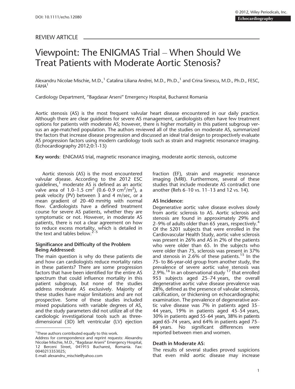
© 2012, Wiley Periodicals, Inc. Echocardiography DOI: 10.1111/echo.12080 REVIEW ARTICLE Viewpoint: The ENIGMAS Trial – When Should We Treat Patients with Moderate Aortic Stenosis? Alexandru Nicolae Mischie, M.D.,1Catalina Liliana Andrei, M.D., Ph.D.,1and Crina Sinescu, M.D., Ph.D., FESC, FAHA1 Cardiology Department, “Bagdasar Arseni” Emergency Hospital, Bucharest Romania Aortic stenosis (AS) is the most frequent valvular heart disease encountered in our daily practice. Although there are clear guidelines for severe AS management, cardiologists often have few treatment options for patients with moderate AS; however, there is higher mortality in this patient subgroup ver- sus an age-matched population. The authors reviewed all of the studies on moderate AS, summarized the factors that increase disease progression and discussed an ideal trial design to prospectively evaluate AS progression factors using modern cardiology tools such as strain and magnetic resonance imaging. (Echocardiography 2012;0:1-13) Key words: ENIGMAS trial, magnetic resonance imaging, moderate aortic stenosis, outcome Aortic stenosis (AS) is the most encountered valvular disease. According to the 2012 ESC guidelines,1moderate AS is defined as an aortic valve area of 1.0–1.5 cm2(0.6–0.9 cm2/m2), a peak velocity (PV) between 3 and 4 m/sec, or a mean gradient of 20–40 mmHg with normal flow. Cardiologists have a defined treatment course for severe AS patients, whether they are symptomatic or not. However, in moderate AS patients, there is not a clear agreement on how to reduce excess mortality, which is detailed in the text and tables below.2–5 fraction (EF), strain and magnetic resonance imaging (MRI). Furthermore, several of these studies that include moderate AS contradict one another (Refs 6–10 vs. 11–13 and 12 vs. 14). AS Incidence: Degenerative aortic valve disease evolves slowly from aortic sclerosis to AS. Aortic sclerosis and stenosis are found in approximately 29% and 2–9% of adults older than 65 years, respectively.4 Of the 5201 subjects that were enrolled in the Cardiovascular Health Study, aortic valve sclerosis was present in 26% and AS in 2% of the patients who were older than 65. In the subjects who were older than 75, sclerosis was present in 37% and stenosis in 2.6% of these patients.15In the 75- to 86-year-old group from another study, the prevalence of severe aortic valve stenosis was 2.9%.16In an observational study17that enrolled 953 subjects aged 25–74 years, the overall degenerative aortic valve disease prevalence was 28%, defined as the presence of valvular sclerosis, calcification, or thickening on echocardiographic examination. The prevalence of degenerative aor- tic valve disease was 7% in patients aged 35– 44 years, 19% in patients aged 45–54 years, 30% in patients aged 55–64 years, 38% in patients aged 65–74 years, and 64% in patients aged 75– 84 years. No significant reported between men and women. Significance and Difficulty of the Problem Being Addressed: The main question is why do these patients die and how can cardiologists reduce mortality rates in these patients? There are some progression factors that have been identified for the entire AS spectrum that could influence mortality in this patient subgroup, but none of the studies address moderate AS exclusively. Majority of these studies have major limitations and are not prospective. Some of these studies included mixed populations with variable degrees of AS, and the study parameters did not utilize all of the cardiologic investigational tools such as three- dimensional (3D) left ventricular (LV) ejection differences were 1These authors contributed equally to this work. Address for correspondence and reprint requests: Alexandru Nicolae Mischie, M.D., “Bagdasar Arseni” Emergency Hospital, 12 Berceni Street, 041915 0040213353025; E-mail: alexandru_mischie@yahoo.com Death in Moderate AS: The results of several studies proved suspicions that even mild aortic disease may increase Bucharest, Romania. Fax: 1
Mischie, et al. mortality.2People with mild or moderate AS and aortic sclerosis have an increased death rate com- pared with an age-matched healthy population. In one study,3asymptomatic adults with moder- ate-to-severe AS were studied at baseline; the authors found a cardiovascular-associated death rate of 3.5% at a mean follow-up (FU) of 2.5 years (4 symptomatic patients died). In 1999, the results of a prospective study4with a 5-year FU were published; a cardiac-associated death rate of 6.1% was found in the general pop- ulation with normal aortic valves, a 10.1% death rate was found in patients with aortic sclerosis, and a 19.6% death rate was found in AS patients. Although the mean age of study patients was 73 years and there was no reported aortic valve surgery (AVS), this study was relevant for the general population that has various risk factors and is also relevant to our review. There is a 4% increase in cardiac-associated death for aortic sclerosis patients and increase in cardiac-associated death for AS patients. In 2004, Rosenhek et al.5reported a cardiac death rate of 8.7% in a population with moderate AS and a mean age of 67 years at the initial examination. Nineteen percent of patients (n = 33) had AVS for severe AS at the 48-month FU. The largest study11evaluating medical treat- ment in moderate AS patients reported a cardiac death rate of 6.0%, which was similar to the gen- eral population; however, the authors considered this study to not be significant for death rate in moderate AS patients because a super-selected population was enrolled (among exclusion crite- ria were coronary artery disease (CAD) history, stroke, and diabetes mellitus). The exact death rates and event-free survival can be found in the tables below. progression, outcomes, survival, limitations, and conclusions. Even though all of the essential moderate AS data can be easily read in the tables, the authors would like to briefly discuss the studies that refer to echocardiography as a tool for stratifying outcomes, and also to debate whether medical interventions had any regression. Monin et al. enrolled 107 patients with a baseline PV of 3.5–4.4 m/sec. Seventy-two per- cent of these patients had severe AS, of which 62 became symptomatic at 2 years.35He concluded that the female sex, PV, and brain natriuretic peptide (BNP) at baseline were correlated with AS progression and developed a score to best stratify outcomes in these patients. Twenty- month survival was 80% for the patients within the first score quartile versus 7% for the patients within the fourth score quartile. A landmark trial, the study of Marechaux et al.36included 135 patients with a mean age of 64 years with mod- erate or severe AS (mean PV of 3.8 ? 0.8 m/sec, 53% had severe AS) and with a normal stress test at the baseline, from which 67 had an event at 20 months. He found that an age of ?65 years, diabetes, LV hypertrophy, resting mean gradient >35 mmHg, and an exercise-induced mean gra- dient increase of >20 mmHg (by multivariate analysis) were independent predictors of death and AVS. Increased progression was present in those patients with a resting mean gradient of >35 mmHg and an exercise-induced mean gradi- ent increase of >20 mmHg. Although this was a remarkable study, it lacks strain analysis, 3D LVEF, twist and torsion; furthermore, an MRI was not performed for fibrosis evaluation. Moreover, the included population was not limited to moderate AS patients. A recent substudy from the SEAS trial that was not included in the tables described that increased left atrial systolic force is a marker of increased cardiovascular events at 4.3 years FU.37Although cardiologists become more and more familiar with strain techniques38with time, few studies clarified the role of strain in AS. One of the studies demonstrated that despite an unchanged LVEF, strain gradually decreased as AS severity increased; therefore, global longitudi- nal strain might be useful to assess subtle changes in LV function in mild, moderate, and severe AS patients.26However, there are impor- tant limitations: this was a nonprospective inves- tigation, only longitudinal strain was measured, there was a small sample size, the groups were nonheterogenic, and reported. The second study by Ng et al.28evalu- ated the systolic function of 420 patients with aortic sclerosis and stenosis with a mean age of 61 years and a normal EF. The author found that impact in disease an additional 13.5% Trials Involving Patients with Moderate AS: In this presentation, the authors will not review or discuss severe AS outcomes and will only dis- cuss moderate AS patient outcomes. The first step in our research was identifying all of the moderate AS studies. The large majority of studies included moderate AS among the AS patient spectrum of mild, moderate, and severe sclerosis, either alone or combined. For that rea- son, the authors of this review categorized stud- ies by the baseline inclusion criteria. We found 4 major groups of enrolled patients as reviewed and discussed below. The 4 groups were patients with mild-to-moderate AS (Table I),5,11,12,18–20 patients with moderate patients with mild, moderate, and severe AS (Table III)14,23–29and patients with moderate-to- severe AS (Table IV).30–36All of the tables repro- duce the hallmark findings from each study, with emphasis on patient characteristics, AS (Table II),3,21,22 AS no progression was 2
The ENIGMAS Trial (continued) events related to Conclusions, Survival older was more moderate AS in events, but not rapid than that 3 and 5 years. of smoking are m/sec and age 60 ? 4% at 1, was 95 ? 2%, cardiovascular and ezetimibe 75 ? 3% and of AS, defined than 80 years patients aged in those aged Mortality was calcifications, Rapid progression with baseline Event free survival 80 years and in those with independent BMI and a history predictors of incidence of events, with mmHg/year reduced the 80% higher progression population than in the with CAD, significant grade 3–4 >50 years Progression of mild and ischemic younger general PV > 3 Simvastatin as >5 patients enrolled cancer in active Lack of onset of (known CAD, symptoms as arm, low-risk High incidence of Limitations stroke, DM Not all had excluded) endpoint Retrospective Retrospective echo FU NA of deaths and AVR Similar percentages outcome was To identify clinical predictors of a composite progression Endpoint Death (n = 34) (n = 33) of MACE The primary or AVR of AS NA calcification, CAD and peak jet velocity were with a progression of Predictors of Outcome aortic-valve stenosis ezetimibe reduced greater: history of 5 mm Hg/year or factors associated events related to the incidence of Independent clinical events, but not outcome (MA) Moderate to severe cardiovascular increased BMI smoking and independent predictors of aortic valve Simvastatin and Trials with Mild-to-Moderate AS as Baseline Inclusion Criteria ischemic Age year If Not Specified Progression (m/sec/ ?0.05 ? 0.06 cm2/year ?0.10 ? 0.08 cm2/year (no event vs. event) Rate of AVA degression: (<50 years vs. >50 6.3 ? 13 mm Hg/year (no CAD vs. CAD) (grade 3–4 calcif. 0.15 ? 0.01 (placebo Otherwise) vs. 0.30 ? 0.33 0.24 ? 0.30 (overall (group >80 years) (group <80 years) vs. grade 1–2) mean rate of years of age) 0.45 ? 0.38 0.35 ? 0.31 0.34 ? 0.42 (Simvastatin + Ezetimibe population) progression 0.16 ? 0.01 0.14 ? 0.18 vs. 0.16 ? 0.19 vs. 0.18 ? 0.19 vs. 0.10 ? 0.14 group) group) TABLE I 48 ? 19 months 52.2 months FU 2.5 years Simvastatin + Ezetimibe of 3.09 ? 0.55 m/sec) Baseline Aortic Valve of 3.1 ? 0.54 m/sec) 3.06 ? 0.38 m/sec) group (n = 944, PV Events group (n = 109, PV of 3.25 ? 0.37 61% with mild and (n = 67, PV of (n = 929, PV Mild-to-moderate moderate AS No events group 31% with Placebo group m/sec) 70.7 ? 10 years 58 ? 19 years 58 ? 19 years Age 75 years asymptomatic AS (mean of ezetimibe or placebo. of simvastatin + 10 mg with mild-to-moderate divided into no events randomized to 40 mg patients (73 women) 3.13 ? 0.39 m/sec) and 21 patients <80 of 84 vs. 66 years) mild-to-moderate, AVA of 1.28 cm2) and events group 87 Patients, 81% men 19 Patients >80 years years (mean age AS (mean PV of 176 Asymptomatic Patients 1873 Patients with Author/Parameter (SEAS trial)11 Rosenhek et al.5 Prospective. Rossebo et al. Kume et al.19 Ngo et al.18 3
Mischie, et al. (35.3% vs. 38.2%, BMI = body mass index; PV = peak velocity; CAD = coronary artery disease; AVA = aortic valve area; NA = nonavailable; AVR = aortic valve replacement; FU = follow-up; DM = diabetes mellitus; AS = aortic stenosis; CRP = C-reactive protein; MACE = major adverse cardiovascular events; MA = multivariate analysis; TDI = tissue Doppler imaging; LA = left atrium; 10.8% vs. 11.1%, mild-to-moderate levels, but has no progression, and deaths, and AVR Conclusions, Survival progression and of AS. CRP does in the placebo Similar percentages and 29.9% vs. progression of clinical events -moderate AS attenuate the patients with effect on the reduces CRP with mild-to prognosis in rosuvastatin dysfunction aortic-valve not predict Treatment with stenosis in of MACE, vs. active diastolic severity, patients Rosuvastatin 28.3%) did not group AS TDI is angle dependent. strain rate, and Vp were not available Small sample size volumes, strain, Medial TDI, LA Limitations Observational Observational of high-sensitivity interaction with the progression rosuvastatin on To examine the role Endpoint CRP and its of AS. NA Age, female gender, Predictors of Outcome -density lipoprotein BMI, and low high progression (MA). cholesterol were associated with elevated CRP. PV predicted AS NA different between patients No difference in progression 4.4 ? 0.5 m/sec at FU. rosuvastatin and those year If Not Specified Progression of AS was not treated with placebo Progression (m/sec/ vs. 3.3 ? 0.7 m/sec in all tertiles of CRP Group 2: 3.2 ? 0.1 at Group 3: 3.7 ? 0.2 at vs. 3.9 ? 0.7 m/ Otherwise) Group 1: 2.8 ? 0.1 for placebo vs. Table I (continued) rosuvastatin: treated with baseline vs. at baseline sec at FU baseline at FU FU 3.5 years 3.5 years placebo vs. rosuvastatin in all Group III: 3.6–4.0 m/sec, Group II: 3.1–3.5 m/sec; Baseline Aortic Valve range 2.5–4.0 m/sec) Group I: 2.5–3.0 m/sec; (mean 3.1 m/sec, Mild-to-moderate AS 56 ? 13 years Age 57 years Vp = propagation speed into left ventricle. double-blind, placebo vs. Rosuvastatin 40 mg. 250 Patients randomized Patients 168 Patients (ASTRONOMER (ASTRONOMER Author/Parameter study)20 study)12 Chan et al. Jassal et al. 4
The ENIGMAS Trial longitudinal, radial, and circumferential strain as well as strain rate deteriorated with aortic valve disease progression. Several limitations may be noted: the study was not prospective; thus, there were no progression numbers, the time to symp- toms was not specified, twist or torsion were not evaluated, clinical variables were not evaluated, and the measurements were performed on differ- ent instruments. Strain was already utilized in severe AS patients; however, those patients are not our tar- get group. Until April 2012, there were no MRI studies on moderate AS patients. The authors of the above study did not dis- cuss very old studies or very small series of AS patients’ studies (most of these are found in Ref. 30). Their capability to address this subject was limited by a retrospective design in most cases, potential selection bias, and limited clinical, func- tional, or exercise data. AS Pathophysiology: Aortic stenosis often progresses slowly over a per- iod of years. During this period of pressure over- load, the LV adapts by sarcomere replication. This remodeling leads to development of con- centric hypertrophy and an increase in LV wall thickness with normal chamber volume. This is enough to counterbalance the increased LV pres- sure and thus preserve LVEF in the initial stages.42 Once these physiological surpassed, chronic pressure overload develops and leads to a depressed LVEF because of impro- per ventricular hypertrophy in response to high LV pressure. Depressed LVEF may also occur because of true myocardial contractility depres- sion, which is explained by alterations in myocar- dial perfusion in the absence of CAD ischemia due to increased LV mass. Finally, depressed LVEF may occur because of a pro- longed ejection period and fibrosis, which often begins in the subendocardium.44,45 AS Progression: There is a wide variability in AS progression, and each study reported differing AS progression rates depending on the FU interval, comorbidi- ties, and patient age (see Tables I–IV). For exam- ple, peak jet velocity rates were as low as 0.15 ? 0.01 m/sec per year in the population without cardiovascular increased to 0.61 ? 0.32 m/sec per year in pri- mary care patients.29 High progressors had at least one of the fol- lowing: mechanisms are factors,11 risk and 43and 1 Echocardiography baseline increased increased mean gradient,3high rate of increase in jet velocity,3,29moderate-to- severe aortic valve calcification on echo,5,22 LV hypertrophy,36resting mean gradient >35 mmHg,36exercise-induced mean gra- dient increase >20 mmHg,36increased E velocity,25bicuspid aortic valve.25 2 Clinical parameters: CAD,5,30 years19, 22, 27, 29, >64 years30, >65 years36), diabetes,23,36metabolic syndrome,33dialy- sis,39,40increased body mass index (BMI),18 functional status,3history of smoking,15,18 systolic blood pressure (SBP),15,27 gender,15,33female gender35; 3 Biological parameters: increased parathy- roid hormone levels,27increased baseline BNP,35increased C-reactive protein levels (>0.15 mg/dL),14high lipoprotein(a) and low-density lipoprotein cholesterol levels.15 parameters: velocity,3,5,12,22,25,29,35 baseline peak increased jet gradient,33 Future Directions: The question in the title is not rhetorical. Because the mortality rate in moderate AS patients is increased, there should be a specific treatment window when medical or surgical interventions for these valvular heart disease patients would be of benefit. Cardiologists should not wait until moderate AS becomes severe, but should aim to nullify the effects of factors that increase mortal- ity and determine whether improve after targeted medical treatment or surgery. Although some progression factors in all AS spectrums may influence mortality, none of the previous studies addressed moderate AS exclusively. Most of these previous studies have major limitations. Because the mortality rate is double or nearly triple in older AS patients com- pared with an age-matched population, future studies must clearly identify and stratify progres- sion risk factors in moderate AS patients for appropriate treatment. A more integrated approach in moderate AS patient management would be to develop a risk score by identifying the clinical parameters, echo- cardiographic parameters (3D, strain, strain rate, twist or torsion at rest and during stress), and MRI parameters (fibrosis extent, LV mass, EF, etc. age (>80 these patients male Treatment with simvastatin and ezetimibe,11 eplerenone34rosuvastatin,20or other statins23 had no influence on AS progression. Slower AS progression patients with osteoporosis21or bisphosphonate treatment,24,41but these studies were too small, retrospective,21,24or biased.21 was observed in 5
Mischie, et al. 2 years and 34 ? 15% PV = peak velocity; AVA = aortic valve area; AVR = aortic valve replacement; FU = follow-up; AS = aortic stenosis; NA = nonavailable; OT = osteoporosis treatment (bisphosphonates, and AV calcifications Conclusions, Survival year, 67 ? 10% at was 93 ? 5% at 1 baseline, the rate of hemodynamic slow progressors clinical outcome progression and the baseline PV, moderate AS at associated with related to age, independently and functional than expected The progression of asymptomatic Survival for overall OT is strongly and AS was slower by jet PV, rate Event-free survival 87.5 ? 8.3% are predicted of change in respectively, progression population jet velocity rate at FU: and it was for fast vs. decreased vs. 100%, at 3 years In adults with status of AS Nonrandomized, Retrospective population, substantial Limitations overlap mixed Biased NA events (death or Death (n = 3.5) or AVR (n = 39) and cardiac Endpoint Progression AVR) NA Predictors of Outcome baseline, the rate functional status of change in jet velocity, and moderate-to incidence of calcification progression progression Baseline PV and score (MA) -severe AV OT associated related to Jet velocity at with AS (MA) Trials with Moderate AS as Baseline Inclusion Criteria If Not Specified Otherwise) Progression (m/sec/year ?0.22 ? 0.22 cm2in ?0.10 ? 0.18 cm2in (slow progressors) (no events group) (fast progressors) AVA regression was: 0.45 ? 0.42 m/sec 0.32 ? 0.34 m/sec 0.23 ? 0.22 m/sec (events group) those not on OT patients on OT (overall) TABLE II 2.2 ? 0.3 2.6 ? 0.3 2.5 ? 1.4 years 2.4 ? 1.0 years FU 6.0 years calcitonin, or estrogen receptor modulators); MA = multivariate analysis. PV of 3.3 ? 0.5 m/sec) Baseline Aortic Valve cm2(1.29 ? 0.46 jet velocity ?2.0 and <3.0 m/sec) vs. 1.39 ? 0.42 Mild AS (peak aortic PV of 3.9 ? 0.5 -asymptomatic cm2-OT group AVA was 1.33 cm2-controls) -AS (n = 67, AS (n = 56, Mean baseline Events groups m/sec) group No events 62.1 ? 11.9 years 63 ? 16 years Age 82 years AVA of 1.3 ? 0.5 Korean patients, PV of 3.6 ? 0.6 and 37 patients 123 Asymptomatic 153 Asymptomatic patients, 70% 18 Patients on OT m/sec, mean moderate AS males, mean not on OT Patients 31 males cm2 Author/Parameter Skolnick et al.21 Prospective Otto et al.3 Seo et al.22 6
The ENIGMAS Trial (continued) with asymptomatic AS associated with slower Bisphosphonate treatment than that reported in prognosis in patients moderate calcific AS Statins do not affect Survival was lower in the Western population LVEF, GLS gradually progression of mild to congestive heart was independently failure, and 13 AVR) AS in patients with hospitalization due (?23 cardiac), 25 faster in DM than patients is slower progression, and CRP predicts severity, high CRP group The progression rate preserved renal Conclusions, of AS in Korean Despite unchanged progression of Calcific AS severity Survival severity of AS subjects with decreased as at baseline. in non-DM (33 deaths progresses calcific AS function Observational. Only long. strain Small size, measured. Retrospective Retrospective Retrospective Retrospective Limitations CRP influences Endpoint Assess whether AS progression AS NA NA NA biophosphonates Trials with Mild, Moderate and Severe AS as Baseline Inclusion Criteria GLS was significantly Progression inversely correlated with correlated with CRP (>0.15 mg/dL) velocity (MA) independent Diabetes predicts of severe AS aortic valve, progression in the rapid E’, LV mass AVA, LVEF, treatment calcific AS predictor bicuspid severity of Outcome Baseline PV, was an CRP higher group and E Predictors groups (mild: 17.1 ? 3.0%, (mean progression rate) – biophosphonates differences among GLS showed significant <0.15 cm2/year-slow (n = 25), Degression Specified Otherwise) Hg/year controls non-DM group (m/sec/year If Not (moderate AS) in bicuspid vs. Degression of AVA moderate AS 0.11 ? 0.20 mmHg/year tricuspid AS cm2/year in cm2/year in m/sec/year 0.23 ? 0.35 vs. (severe AS) DM group ?0.14 ? 0.13 ?0.25 ? 0.20 2.8 ? 3.3 mm cm2/year-fast of AVA >0.15 Progression in (mild AS) 0.28 ? 0.36 0.14 ? 0.26 0.09 ? 0.18 progressors progressors 0.12 ? 0.23 TABLE III Progression group: group 0.1 ? 3.3 the 3 (n = 22) No FU, observational Mild AS: 2.93 years Severe AS: 1.69 Moderate AS: 29 ? 13 months 23 ? 11 months 2003–2008 2.40 years Retrospective, FU years and severe in 25 patients. Mild (PV of 2.7 ? 0.3 m/sec), (PV of 3.7 ? 0.4 m/sec), Mild AS (n = 18, 71 ? 10 years), Moderate AS Severe AS (n = 60, (25%), and severe (n = 57, 77 ? 9), (64%), moderate AS in 37 patients 22 had mild and Aortic Valve group (n = 28), moderate in 75 Calcific AS was mild 6 moderate-to Baseline in 66 subjects, Bisphosphonates and severe -severe AS Mild AS in 207 moderate of whom AS in 81 78 ? 8) (11%) 73.3 ? 8.8 years 68 ? 10 years 67 ? 13 years 70 ? 9 years Age 76 years Moderate AS (n = 25), 113 Patients, 38% males Severe AS (n = 39) Mild AS (n = 49), which 72 (43%) 46 moderate-to 325 Korean patients preserved renal 57 had mild AS and calcific AS, of 166 Patients with 103 Patients with Patients -severe AS function had DM 135 Patients Sterbakova et al.24 Author/Parameter Kamalesh et al.23 Miyazaki et al.26 Imai et al.14 Ryu et al.25 7
Mischie, et al. (continued) elderly and frequently the subendocardium with asymptomatic with asymptomatic severity (by strain). with normal LVEF, appears to start in severe AS patients high PTH and SBP in the presence of and progresses to predicts, not only dysfunction with In aortic valve disease dysfunction that in LV function in strain compared had a decreased Primary care patients overall mortality Conclusions, independently hemodynamic AS progression was moderate and develop rapid GLS might be AS are usually increasing AS Survival AVR, but also hemodialysis with chronic assess subtle progression, accelerated AS patients myocardial transmural in patients increased. there is a Symptomatic useful to changes patients patients which of MI excluded, nonheterogenic. randomized, no Blood pressure obtained from with WMA or not measured, to symptoms Not prospective, progression torsion/time no twist/ Retrospective Retrospective reported, patients records Limitations history not patients with AS To define risk factors multidirectional strain and SR in progression in mortality and AVR Identify changes in All-cause mortality hemodialysis and a composite affecting AS underwent AVR 40 died and 48 Endpoint patients chronic of all-cause the composite endpoint were the yearly change The independent predictors in PV and age (MA); on the initial echo the yearly change of death and AVR of mortality were The predictors of associated with independently hypertension in PV and PV progression index, and associated LVEF, and GLS (MA) SBP (MA) PTH level and with AS of Outcome MPG, Predictors were NA ?18.0 ? 1.7, ?17.1 ? 2.0, ?21.1 ? 3.7, ?19.7 ? 3.3, 50.3 ? 17.5, 47.4 ? 13.2, Long. strain: ?20.3 ? 1.9, Radial strain: 53.7 ? 14.8, and severe: 14.5 ? 3.9%, Circ. strain: ?22.2 ? 3.3, moderate: 16.4 ? 3.0%, following (from sclerosis progression, 49 patients (slow progression, 104 0.29 ? 0.19 cm2/year 0.03 ? 0.02 cm2/year group, 20 patients) group, 14 patients) for the 4 groups as /mild/moderate to (rapid progression (slow progression Table III (continued) Specified Otherwise) Strain (%) decreased patients [68%]) (m/sec/year If Not ?15.1 ? 2.4 ?17.9 ? 4.0 AVA reduction of: 0.61 ? 0.32 (fast 41.1 ? 15.7 severe AS): P = 0.003) 0.10 ? 0.16 [32%]) Progression (rapid progressors) (slow progressors) 4.9 ? 2.7 years 20 ? 9 months months FU 26 ? 8 No FU (PV of 4.9 ? 0.5 m/sec) (n = 20), and severe AS AVA of 1.0 ? 0.3 cm2 Aortic sclerosis (n = 118), of 2.7 ? 0.56 m/sec Mild (n = 9), moderate 1.31 ? 0.31 cm2 mild AS (n = 81), (n = 109), severe 3.2 ? 0.6 m/sec of moderate and Aortic Valve (n = 5); AVA of Symptoms in 58.7% mild, 71 (46%) Baseline and 18 (12%) AS with mean and mean PV AS (n = 112) moderate AS 135 patients with mean PV of (42%) had moderate, severe AS severe AS 64 patients 66.1 ? 14.5 years 69 ? 8 years 77 ? 9 years Age had multidirectional imaging performed patients, 65% men men with aortic sclerosis and AS 420 Patients, 60.7% 153 Asymptomatic strain and SR hemodialysis Patients patients 34 Chronic Author/Parameter Iwata et al.27 Nistri et al.29 Ng et al.28 8
The ENIGMAS Trial at rest and stress) that expose a portion of fast-progressing moderate AS patients to a higher rate of events than the slow-progressing moder- ate AS patients. The ENIGMAS trial, a prospective study focused on identification of clinical, biological and imagistic parameters in rapid-progression subgroup patients with moderate aortic stenosis, is divided into a prospective observational study and a prospective interventional study, and seems to be an ideal trial for patient evaluation (ClinicalTrials.gov no. NCT01575249). The study protocol is highlighted in Fig. 1. The ENIGMAS trial will start enrolling patients in July 2012, will last for 3 years, and will include only moderate AS patients. It will include symptomatic and asymptomatic moderate AS patients and will add new evaluation techniques such as newer echo parameters, strain and twist, and MRI. Mild AS patients with an initial PV of 2.8–3.1 m/sec will be divided into 2 groups. The first (observa- tional) group will have 160 patients with a nega- tive exercise stress echo for symptoms/ECG/wall- motion abnormalities (WMA), a negative spirom- etry test for pulmonary disease, no known CAD or other valvular diseases, and an in-sinus rhythm and an LVEF > 55%. The second group (medi- cal/surgery group) will also have 160 patients with symptomatic AS as determined by negative pulmonary tests but positive stress echo, prior CAD or other valvular LVEF > 55%). One major difference of this study from other studies is that the echo exams will be performed on identical because strain is instrument dependent. Patients will be followed every 6 months for 3 years with clinical, biological, and stress echo exams. MRI will also be included at each FU to evaluate LV mass, LVEF, WMA, calcifications, and fibrosis so that this moderate AS patient subgroup will have complete cardiologic data for evaluation. The primary endpoint will be the occurrence of major adverse cardiac events defined by the following: death and AVS. The secondary endpoint will be the time that it takes to develop stress symptoms such as angina, dyspnea, or syncope in the first group. The tertiary endpoint will be the time that it takes to develop altered stress-related hemody- namic parameters in both groups such as strain decrease, EF decrease, ventricular arrhythmia, SBP decrease, and pathological stress gradients. The role of fibrosis and other MRI manifestations of pathological changes in moderate AS patients will also be clarified. Investigators will also differ- entiate patient outcomes with mean gradients between 40 and 50 mmHg, which is the gray AS zone, because currently there is a discrepancy between the American and European guidelines for the severe AS cutoff.1,46 rapid hemodynamic AS at baseline and mild-to-moderate Similar event-free Conclusions, with severe AS. and in patients Survival patients with progression Survival worse in major adverse cardiovascular events; GLS = global longitudinal strain; LVEF = left ventricular ejection fraction; MPG = mean peak gradient; WMA = wall-motion abnormalities; survival PV = peak velocity; AVA = aortic valve area; AVR = aortic valve replacement; FU = follow-up; DM = diabetes mellitus; AS = aortic stenosis; CRP = C reactive protein; MACE = Limitations Endpoint of Outcome Predictors Table III (continued) Specified Otherwise) (m/sec/year If Not Progression diseases, and an MI = myocardial infarction; MA = multivariate analysis; SBP, systolic blood pressure. echo instruments FU Aortic Valve Baseline Age Patients Author/Parameter 9
Mischie, et al. (continued) The PPV for exercise-induced symptoms was 57% in 49% for symptomatic the whole population the whole population and 79% for patients and worse outcome The NPV was 87% in identify a population Activity Scale Class I. 10.6 ? 11.0 mmHg There was no AVR. Nearly half the patients progression/year of MS is associated with a in patients with AS on exercise testing -free survival at 12 severe AS reveal a Conclusions, Survival asymptomatic. faster disease the subgroup. An abnormal test symptoms or of ?10 mm moderate to for closer FU with initially progression progression and 86% in versus 89% year. Mean may reveal in Specific Symptom aged <70 for those months: patients Hg per defined (severe AVA < 0.8 cm2) AS not correctly Limitations Retrospective. Small study AS: – symptoms (n = 36) or death (n = 0) at FU and AVR (n = 45) (n = 9 vs. n = 4), death (n = 3 vs. n = 1) in rapid cardiovascular cardiac, n = 3 Endpoint spontaneous Development of Symptoms, AVR noncardiac) AVR (n = 16) exertional Death (n = 5 progress vs. slow and event-free survival (MA) gradient were independent MS, males and baseline peak predictors of progression Predictors of Outcome symptoms. At FU, 10 Increased progression in normal exercise ST older patients (64 Trials with Moderate-to-Severe AS as Baseline Inclusion Criteria as they remained abnormal ST did not require AVR patients with a asymptomatic and with CAD and 4 with an (38% vs. 7%) (PPV of 78%) Exercise-limiting Exercise-limiting vs. 53 years) symptoms normal and abnormal exercise tests There was no statistically significant and MG between patients with difference in AV area, maximal, Not Specified Otherwise) Progression (m/sec/year If had a PV of 4.1 ? 0.6 m/sec (rapid progressors, n = 21), 4 ? 3 mmHg increase/year patients with no end-point 19 ? 12 mmHg increase/year (slow progressors, n = 28) and ?0.14 ? 0.13 cm2/year Patients with endpoint versus 0.19 ? 0.27 (all population), versus 3.7 ? 0.8 m/sec 0.28 ? 0.30 m/sec/year 0.13 ? 0.24 m/sec/year TABLE IV (MS patients) NA 32 ? 16 months 28 ? 13 months 12 months 36 months FU baseline AVA: 1.08 ? 0.24 cm2, Baseline PV: 3.2 ? 0.6 m/sec, Baseline Aortic Valve than 42% had severe AS Moderate (n = 20) and 40% had severe AS severe (n = 10) AS AVA < 1.4 cm2, more 38 ? 15 mmHg 62 ? 14 years 69 ? 12 years 58 ? 16 years Age 65 years progressors at FU symptoms at FU from which 36 30 Asymptomatic spontaneous MS, 64 men Patients from which 49 Patients, 29 were rapid developed 38% had males, 21 125 Patients, 105 Patients, patients Author/Parameter Alborino et al.31 Briand et al.33 Peter et al.30 Das et al.32 10
The ENIGMAS Trial AS = aortic stenosis; PV = peak velocity; AVA = aortic valve area; AVR = aortic valve replacement; FU = follow-up; CAD = coronary artery disease; NA = not available; MS = metabolic syndrome; MA = multivariate analysis; LV = left ventricle; ST = stress test; ESE = exercise stress echocardiography; MG = mean gradient; PPV = positive predictive value; NPV = negative (44 ? 8% vs. 69 ? 6%) eplerenone did not slow or diastolic dysfunction, true asymptomatic AS the onset of LV systolic predictors of outcome reduce progression of decrease LV mass, or ESE, but not indices moderate-to-severe Hemodynamic indices for patients within with outcome in Conclusions, Survival Authors developed a exercise capacity on independent Survival after 20 among patients risk score based are associated quartile vs. 7% aortic stenosis, for the fourth the first score patients with valve stenosis. measured by of maximum months: 80% In asymptomatic patients with Similar survival Survival lower with MS quartile measurements, 72% had severe Limitations Study too small AS, 22.1% for clinical outcomes had CAD no strain No BNP systolic dysfunction due to symptoms due to symptoms or LV dysfunction or LV hypertrophy Eplerenone effect on in asymptomatic refused surgery or positive ST, AVR (n = 58) AVR (n = 59) Endpoint patients with Death (n = 3) or Death (n = 3) or moderate to delay of LV 1 patient severe AS Predictors of Outcome -induced increase MG >35 mmHg, LV hypertrophy, BNP at baseline Age ?65, diabetes, Female sex, PV and gradient >20 mmHg (MA) and exercise in mean resting None 0.4 m/sec in symptomatic patients Not Specified Otherwise) Table IV (continued) Progression (m/sec/year If vs ?0.18 ? 0.24 cm2/year and ?0.08 ? 0.08 cm2/year for elprenone vs. placebo 0.2 m/sec in asymptomatic, MG >35 mmHg and gradient >20 mmHg Similar decrease of AVA Increased progression increase in mean exercise-induced (?0.11 ? 0.22 group, P = ns) in those with resting (controls) 20 ? 14 months 19 months 24 months FU baseline PV of 3.92 m/sec /sec or placebo (n = 32) Baseline Aortic Valve baseline PV of 3.85 m Mean PV of 3.8 ? 0.8 m mg daily (n = 33) Eplerenone group 100 mean 4.1 m/sec /sec, 53% had 3.5–4.4 m/sec, severe AS 64 ? 15 years 67.5 years Age 72 years patients randomized 135 Patients with at least symptomatic at FU moderate AS, with which 67 had an to elprenone or AS, from which normal ST, from 107 Asymptomatic 65 Asymptomatic double-blind Patients 62 became event at FU placebo predictive value. Marechaux et al.36 Author/Parameter Stewart et al.34 Prospective Prospective Monin et al.35 11
Mischie, et al. Design of The ENIGMAS Trial (Prospective study focused on idEntification of cliNical, biological and imagistic parameters in rapid proGression subgroup patients with Moderate Aortic Stenosis) MILD-EARLY MODERATE AS (peak vel. 2.8-3.1 m/s) Exclusion criteria? No Exercise stress echo Positive/Symptoms (interventional group) Negative/No symptoms and Negative Spirometry Negative Spirometry Initial (and FU) evaluation: Coronary angiography Clinical evaluation, blood samples and ECG. Echo parameters at rest and stress (see text for more info): 2D, 3D, strain, LV mass, calcifications. MRI at rest and stress (see text for more info): mass, LVEF, calcifications, fibrosis. CAD Treat as appropriate No CAD RANDOMIZE FU every 6 months (FOR 3 YEARS) AVS Medical treatment PRIMARY ENDPOINT: MACE (death, AVS and myocardial infarction) SECONDARY ENDPOINT: TIME TO SYMPTOMS (angina, dyspnea, syncope) at stress FU every 6 months (FOR 3 YEARS) TERTIARY ENDPOINT: TIME TO ALTERED HEMODINAMIC PARAMETERS at stress (strain decrease, EF decrease, ventricular arrhythmia, decrease in arterial BP, stress gradients suggestive of severe AS) PRIMARY myocardial infarction and aortic prosthesis related major complications) ENDPOINT: MACE (death, QUATERNARY ENDPOINT: differentiate outcomes of patients with mean gradients between 40 and 50 mmHg Figure 1. Design of The ENIGMAS Trial. AS = aortic stenosis; AVS = aortic valve surgery; BP = blood pressure; CAD = coronary artery disease; EF = ejection fraction; FU = follow-up; LV = left ventricle; MACE = major adverse cardiac events; MRI = magnetic resonance imaging. Conclusions: Moderate AS patients have poorer health com- pared with the healthy population. With the use of newly available investigational techniques, finding the parameters that accelerate moderate- to-severe AS will translate into proper medical care or treatment interventions to reduce AS pro- gression, resulting in reduced mortality rates. The ENIGMAS trial is designed to highlight disease progression factors, and perhaps it will provide a substantial benefit to not only patients but also the medical community. In addition, investiga- tors hope to reach a consensus regarding the severe AS cutoff values. echocardiographic, and exercise predictors of outcome. Circulation 1997;95:2262–2270. 9142003. 4. Otto CM, Lind BK, Kitzman DW, et al: Association of aor- tic-valve sclerosis with cardiovascular mortality and mor- bidity in the elderly. N Engl J Med 1999;341:142–147. 5. Rosenhek R, Klaar U, Schemper M, et al: Mild and mod- erate aortic stenosis. Natural history and risk stratification by echocardiography. Eur Heart J 2004;25:199–205. Pub- Med PMID: 14972419. 6. Rajamannan NM, Subramaniam M, Springett M, et al: Atorvastatin inhibits hypercholesterolemia-induced cellu- lar proliferation and bone matrix production in the rabbit aortic valve. Circulation 2002;105:2660–2665. 7. Rosenhek R, Rader F, Loho N, et al: Statins but not angio- tensin-converting enzyme inhibitors delay progression of aortic stenosis. Circulation 2004;110:1291–1295. 8. Novaro GM, Tiong IY, Pearce GL, et al: Effect of hydrox- ymethylglutaryl coenzyme A reductase inhibitors on the progression of calcific 2001;104:2205–2209. 9. Shavelle DM, Takasu J, Budoff MJ, et al: HMG CoA reduc- tase inhibitor (statin) and aortic valve calcium. Lancet 2002;359:1125–1126. 10. Moura LM, Ramos SF, Zamorano JL, et al: Rosuvastatin affecting aortic valve progression of aortic stenosis. J Am Coll Cardiol 2007;49: 554–561. 11. Rossebø AB, Pedersen TR, Boman K, et al; SEAS Investiga- tors: Intensive lipid lowering with simvastatin and ezetim- ibe in aortic stenosis. N Engl J Med 2008;359:1343–1356. 12. Chan KL, Dumesnil JG, Tam J, et al: Effect of rosuvastatin on C-reactive protein and progression of aortic stenosis. Am Heart J 2011;161:1133–1139. PubMed PMID: aortic stenosis. Circulation Acknowledgements: The study has received funding from the Romanian Ministry of Health and the European Commission. References 1. The Joint Task Force on the Management of Valvular Heart Disease of the European Society of Cardiology (ESC) and the European Association for Cardio-Thoracic Surgery (EACTS): Guidelines on the management of valvular heart disease (version 2012). Eur J Cardiothorac Surg 2012;42:S1–S44. 2. Otto CM: Aortic stenosis: Even mild disease is significant. Eur Heart J 2004;25:185–187. 3. Otto CM, Burwash IG, Legget ME, et al: Prospective study of asymptomatic valvular aortic stenosis. Clinical, endothelium to slow the 12
The ENIGMAS Trial 13. Cowell SJ, Newby DE, Prescott RJ, et al: A randomized trial of intensive lipidlowering therapy in calcific aortic stenosis. N Engl J Med 2005;352:2389–2397. 14. Imai K, Okura H, Kume T, et al: C-Reactive protein predicts severity, progression, and prognosis of asymp- tomatic aortic valve stenosis. Am Heart J 2008;156:713– 718. 15. Stewart BF, Siscovick D, Lind BK, et al: Clinical factors associated with calcific aortic valve disease: Cardiovascu- lar Health Study. J Am Coll Cardiol 1997;29:630–634. 16. Lindroos M, Kupari M, Heikkila J, et al: Prevalence of aor- tic valve abnormalities in the elderly: An echocardio- graphic study of a random population sample. J Am Coll Cardiol 1993;21:1220–1225. 17. Stritzke J, Linsel-Nitschke P, Markus MR, et al; MONICA/ KORA Investigators: Association between degenerative aortic valve disease and long-term exposure to cardiovas- cular risk factors: Results of the longitudinal population- based KORA/MONICA survey. Eur Heart J 2009;30:2044– 2053. 18. Ngo MV, Gottdiener JS, Fletcher RD, et al: Smoking and obesity are associated with the progression of aortic ste- nosis. Am J Geriatr Cardiol 2001;10:86–90. 19. Kume T, Kawamoto T, Okura H, et al: Rapid progres- sion of mild to moderate aortic stenosis in patients older than 80 years. J Am Soc Echocardiogr 2007;20: 1243–1246. 20. Jassal DS, Bhagirath KM, Karlstedt E, et al: Evaluating the effectiveness of rosuvastatin in preventing the progres- sion of diastolic dysfunction in aortic stenosis: A substudy of the aortic stenosis progression observation measuring effects of rosuvastatin (ASTRONOMER) study. Cardiovasc Ultrasound 2011;9:5. 21. Skolnick AH, Osranek M, Formica P, et al: Osteoporosis treatment and progression of aortic stenosis. Am J Cardiol 2009;104:122–124. 22. Seo JS, Kang DH, Kim DH, et al: Predictors of echocardio- graphic progression in patients with mild aortic stenosis. Korean Circ J 2011;41:649–653. 23. Kamalesh M, Ng C, El Masry H, et al: Does diabetes accelerate progression of calcific aortic stenosis? Eur J Echocardiogr 2009;10:723–725. 24. Sterbakova G, Vyskocil V, Linhartova K: Bisphosphonates in calcific aortic stenosis: Association with slower progres- sion in mild disease–a pilot retrospective study. Cardiol- ogy 2010;117:184–189. 25. Ryu DR, Park SJ, Han H, et al: Progression rate of aortic valve stenosis in Korean patients. J Cardiovasc Ultrasound 2010;18:127–133. 26. Miyazaki S, Daimon M, Miyazaki T, et al: Global longitu- dinal strain in relation to the severity of aortic stenosis: A two-dimensional speckle-tracking study. Echocardiogra- phy 2011;28:703–708. 27. Iwata S, Hyodo E, Yanagi S, et al: Parathyroid hormone and systolic blood pressure accelerate the progression of aortic valve stenosis in chronic hemodialysis patients. Int J Cardiol 2011 24 June [Epub ahead of print]. 28. Ng AC, Delgado V, Bertini M, et al: Alterations in multidi- rectional myocardial functions in patients with aortic steno- sis and preserved ejection fraction: A two-dimensional speckle tracking analysis. Eur Heart J 2011;32:1542–1550. 29. Nistri S, Faggiano P, Olivotto I, et al: Hemodynamic pro- gression and outcome of asymptomatic aortic stenosis in primary care. Am J Cardiol 2012;109:718–723. 30. Peter M, Hoffmann A, Parker C, et al: Progression of aor- tic stenosis. Role of age and concomitant coronary artery disease. Chest 1993;103:1715–1719. 31. Alborino D, Hoffmann JL, Fournet PC, et al: Value of exer- cise testing to evaluate the indication for surgery in asymptomatic patients with valvular aortic stenosis. J Heart Valve Dis 2002;11:204–209. 32. Das P, Rimington H, Chambers J: Exercise testing to stratify risk in aortic stenosis. Eur Heart J 2005;26:1309– 1313. 33. Briand M, Lemieux I, Dumesnil JG, et al: Metabolic syn- drome negatively influences disease progression and prognosis in aortic stenosis. 2006;47:2229–2236. 34. Stewart RA, Kerr AJ, Cowan BR, et al; ZEST Study Investi- gators: A randomized trial of the aldosterone-receptor antagonist eplerenone in asymptomatic moderate-severe aortic stenosis. Am Heart J 2008;156:348–355. 35. Monin JL, Lancellotti P, Monchi M, et al: Risk score for predicting outcome in patients with asymptomatic aortic stenosis. Circulation 2009;120:69–75. 36. Mar? echaux S, Hachicha Z, Bellouin A, et al: Usefulness of exercise-stress echocardiography for risk stratification of true asymptomatic patients with aortic valve stenosis. Eur Heart J 2010;31:1390–1397. 37. Cioffi G, Cramariuc D, Dalsgaard M, et al: Left atrial sys- tolic force and outcome in asymptomatic mild to moder- ate aortic stenosis. Echocardiography 2012;29:1038– 1044. 38. Ammar KA, Paterick TE, Khandheria BK, et al: Myocardial mechanics: Understanding and applying three-dimen- sional speckle tracking echocardiography in clinical prac- tice. Echocardiography 2012;29:861–872. 39. Perkovic V, Hunt D, Griffin SV, et al: Accelerated progres- sion of calcific aortic stenosis in dialysis patients. Nephron Clin Pract 2003;94:c40–c45. 40. Kume T, Kawamoto T, Akasaka T, et al: Rate of progres- sion of valvular aortic stenosis in patients undergoing dialysis. J Am Soc Echocardiogr 2006;19:914–918. 41. Innasimuthu AL, Katz WE: Effect of bisphosphonates on the progression of degenerative aortic stenosis. Echocardi- ography 2011;28:1–7. 42. Wachtell K: Left ventricular systolic performance in asymptomatic aortic stenosis. Eur Heart J Suppl 2008;10: E16–E22. 43. Rajappan K, Rimoldi OE, Dutka DP, et al: Mechanisms of coronary microcirculatory dysfunction in patients with aortic stenosis and angiographically normal coronary arteries. Circulation 2002;105:470–476. 44. Heymans S, Schroen B, Vermeersch P, et al: Increased cardiac expression of tissue inhibitor of metalloprotein- ase-1 and tissue inhibitor of metalloproteinase-2 is related to cardiac fibrosis and dysfunction in the chronic pressure-overloaded human 2005;112:1136–1144. 45. Weidemann F, Herrmann S, Stork S, et al: Impact of myo- cardial fibrosis in patients with symptomatic severe aortic stenosis. Circulation 2009;120:577–584. 46. Castel AL, Mar? echaux S, Laaouaj J, et al: Relationship between cutoff values of peak aortic valve velocity and those of other doppler echocardiographic parameters of severity in patients with aortic stenosis and normal flow. Echocardiography 2012. August 3 [Epub ahead of print]. J Am Coll Cardiol heart. Circulation 13