Download
1 / 39
1.4k likes | 2.92k Views
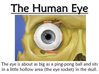
and the Human Eye. Anatomy of the Eye. 1. Sclera. The white part that surrounds the whole eyeball It is a tough, outer protective layer of the eye. Very hard to penetrate. The sclera surrounds the whole eyeball. 2. Cornea.

E N D
1. Sclera • The white part that surrounds the whole eyeball • It is a tough, outer protective layer of the eye. • Very hard to penetrate.
2. Cornea • Curved transparent outer layer where light first enters the eye. • This is where refraction of light first occurs. Cornea
3. Aqueous Humor • Watery fluid which provides pressure to hold the shape of the cornea. Aqueous Humor
4. Pupil • Circular opening in the centre of the eye • The pupil opens up in order to allow more light into the interior of the eye in low light. • The pupil closes up more in order to block some of the light in bright conditions. Pupil
5. Iris • The coloured portion (green, blue etc.) of the eye surrounding the pupil • The iris contains muscles that contract/relax to open/close the pupil. They respond to light levels. At night your pupils are wide open! Iris
6. Lens • Flexible converging lens made of proteins. • Changes shape in order to adjust the focal length for closer or farther objects. (There are muscles and ligaments attached to the lens which stretch the lens.) Lens
7. Vitreous Humor • Liquid/gel-like sac that contains mostly water. • Provides support for the eyeball – keeps the shape of the whole eyeball Vitreous Humor
8. Retina • Thin layer of light-sensitive cells on the back of the eye where the image forms • Two types of photoreceptor cells: - Rods (light and dark sensitive) - Cones (colour sensitive ) Retina
R e t i n a Rods ~ 120 million(light/dark sensitive cells) - 1000 times more sensitive than cones Cones ~6 million(colour sensitive cells) Three types of cone based on colour sensitivity- Red cones “see” red- Green cones “see” green- Blue cones “see” blue
Rod Details • Only see light - not sensitive to color. • Can be triggered by very low light • After 30 minutes at night, our vision gets better as these cells work harder! • Excellent motion sensors. • At nighttime, your eyes are very sensitive to light but cannot discern colours well at all. We see in shades of grey.
9. The Optic Nerve • Carries signals from the cones and rods in the retina to the brain – and we “see” the image! Optic Nerve
Blind Spot • Where the nerve attaches to the retina there are no rods/cones - this region is called the blind spotand each eye contains one.
Image Formation The image is inverted (converging lens!) on the retina and therefore the signal to the brain is of an inverted image. L – Closer O – Inverted (upside down)S – SmallerT - Real The brain gets the upside down signal and flips it so we see things right way up!
Vision Defects • Myopia – near sighted – means you can see things up close, but not focus well on distant objects • Hyperopia – far sighted – can see things far away but cannot focus on close objects (such as reading)
Myopia (nearsightedness) • The eyeball is too long. • The eye focuses light in front of the retina.
Diverging lenses spread the rays apart before entering the eye and focus light onto the retina
Hyperopia (farsightedness) • The eyeball is too short. • The eye focuses light behind the retina.
Converginglenses– cause the rays to converge before entering the eye and focus light on the retina
Astigmatism • The cornea is oval (football shaped) rather than rounded. • Light from objects refract differently because of this difference in curvature • Light focuses at different regions in the eye.
Corrective lenses need to have separate powers for the horizontal (across) and vertical (up and down) defects
Presbyopia • As a person ages, their lens becomes less flexible. The lens cannot change shape as easily • This results in the loss of vision for near objects (the person is now farsighted). • Generally occurs after age 40. • Solution: Reading Glasses – Converging Lenses
If age related presbyopia and myopia are both present – bifocals or progressive lenses can be used.
Laser Eye Surgery • Also called LASIK • Can correct all defects except presbyopia • Uses a laser to reshape the cornea. • Changes the refraction at the cornea so image forms on the retina. • Benefits: No more glasses / contacts! • Risks: Few (now) – dry eyes / infection…..
Age • Laser eye surgery is available only for those age 18 and over. • The shape of the eye is still changing until about age 18. • More importantly the person's eye prescription needs to be stable for at least one year prior to surgery
Video • http://www.youtube.com/watch?v=7PJ391MDtpo • http://www.sciencefriday.com/videos/watch/10059
Sheep’s Eye Dissection • http://www.exploratorium.edu/learning_studio/cow_eye/