Download
1 / 86
1.06k likes | 1.79k Views
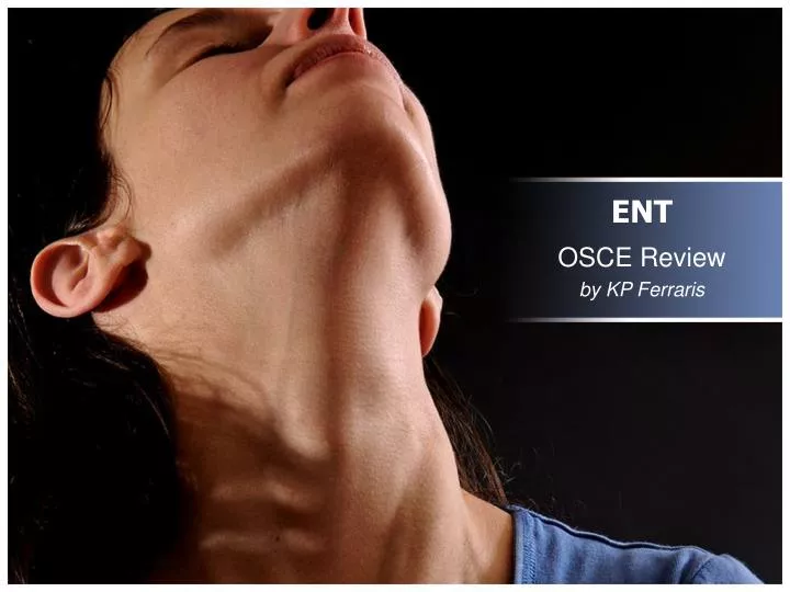
ENT. OSCE Review by KP Ferraris. Outline. 10-pt identification quiz Case presentations with quiz Case 1 Case 2 Case 3 Other ENT symptoms Laundry list of must-know and common diagnoses (For best resolution, view this as Slide Show.). Outline. 10-pt identification quiz

E N D
ENT OSCE Review by KP Ferraris
Outline • 10-pt identification quiz • Case presentations with quiz • Case 1 • Case 2 • Case 3 • Other ENT symptoms • Laundry list of must-know and common diagnoses (For best resolution, view this as Slide Show.)
Outline • 10-pt identification quiz • Case presentations with quiz • Case 1 • Case 2 • Case 3 • Other ENT symptoms • Laundry list of must-know and common diagnoses
Identification Quiz • Shown below is a Lateral X-Ray of the neck showing the thumbprint sign. What is the most likely diagnosis? • Retropharyngeal abscess • Epiglottitis • Acute Tonsillopharyngitis • Croup Answer: b. Epiglottitis (double t in the first only). It is characterized by fever, drooling, dysphagia, odynophagia, noisy breathing, stridor
Identification Quiz 2. Identify the lesion/diagnosis. The larger field is oral mucosa. Answer: Aphthous ulcer or Aphthous stomatitis. It is an erosion of the mucosa caused by either trauma, hot foods/liquid, or lack of hygiene.
Identification Quiz 3. Give the complete diagnosis, with Grading. Answer: Tonsillitis Grade III with Peritonsillar Abscesses (Quinsy). The usual complaint is odynophagia and dysphagia, with or without fever.
Identification Quiz • What type of fracture is shown? • Le Fort I • Le Fort II • Le Fort III • Tripod • Temporal bone Answer: b. Le Fort II fracture. In contradistinction to Le Fort I which is only in the maxilla (upper jaw) and Le Fort III which involves the inferolateral portion of the orbit (cheek).
Identification Quiz 5. Identify this diagnostic test used to confirm Benign Paroxysmal Positional Vertigo (BPPV). Answer: Dix-Hallpike test. It is confirmatory for only one pathology: BPPV, a disorder of the posterior semicircular canal. If positive, where the head is turned to is the side of the lesion.
Identification Quiz 6. Classify the cleft lip and palate using the Thallwitz nomenclature. Answer: L3 A3 H3 S3 H3 A3 L3 (Remember from RIGHT to LEFT, and only one S). Mnemonic is “Lahshal.” This is surgically corrected by Cheiloalveolorhinoplasty.
Identification Quiz 7. Which is the most important part of the sinonasal anatomy commonly blocked during Sinusitis? Answer: Ostiomeatal Unit. It is the common drainage of all sinuses EXCEPT 2: posterior ethmoid cells and the sphenoid sinus.
Identification Quiz 8. What is the most common etiologic agent (bacteria) of Otitis Media? Answer: Streptococcus pneumoniae. H. influenzae is more common in pedia. Another agent is M. catarrhalis.
Identification Quiz • Below is a picture from posterior rhinoscopy. It is the potential site of growth for Nasopharyngeal carcinoma. • Vallecula • Rosenmüller’sfossa • Pyriform sinus • Choana Answer: b. Rosenmüller’s fossa. It is a pharyngeal recess (bilateral) at the back of the nose (nasopharynx) near the torus tobarius surrounding the entrance to the Eustachian tube.
Identification Quiz 10. Interpret the Audiogram below. Answer: Sensorineural hearing loss, mild, AD. See next slide for explanations. AD means right ear (cf. AS, left ear)
Audiogram Remember:Air (AC) uses shapes and X, Bone (BC) uses [ ] and greater-than less-than.
Audiogram Sensorineural hearing loss In sensorineural, both AC and BC are >25 dB. But BC dipped in higher freq. Conductive hearing loss BC is normal while AC is >25 dB. Mixed hearing loss Similar to sensorineural, both AC and BC are >25 dB in mixed. But both AC and BC really dip together.
Outline • 10-pt identification quiz • Case presentations with quiz • Case 1.0, 1.1, 1.2, 1.3, 1.4 • Case 2 • Case 3 • Other ENT symptoms • Laundry list of must-know and common diagnoses
Case 1.0 Hx: OPQRST of Pain, Colds? Allergies? Rode airplane/diving/swimming? Discharge (Otorrhea)? Fever? Hearing loss? Dizziness? Headache? A.Y., a 19 year-old male complained of otalgia. What questions about the history will you ask? What will you perform on P.E.? Inspect for craniofacial anomalies, Otoscopy, Anterior rhinoscopy, Posterior rhinoscopy/Nasal endoscopy, Weber test, Rinne test, Schwabach test (due to the hearing loss) and complete HEENT exam (because may be Referred only).
Case 1.0 2 years PTC:recurrent otorrhea Sought consult, prescribed w/ unrecalled meds (oral and topical drops), doctor said that tympanic membrane was intact. 1 year PTC:otalgia and otorrhea worsened; consult revealed perforated tympanic membrane. 2 weeks PTC: low-grade fever, headache localized to temporal bone, dizziness, persistent otalgia and otorrhea Hx revealed:
Case 1.0 You took your Welch Allyn and Otoscopy revealed: How would you describe the otoscopic findings?
Review of Otoscopy It is important to memorize the anatomic parts of the tympanic membrane to be able to say where the perforation is, where hyperemia is, where the keratin debris are, or where the serosanguinous fluid is coming out. The cone of light reflex (from the reflection of the otoscope light) always points anterior. So this is the RIGHT ear.
Case 1.0 How would you describe the otoscopic findings? Perforation at the pars flaccida, 30% Serosanguinous fluid behind the TM (discoloration) Hyperemia in the Epitympanum
Case 1.0 Pure Tone Audiometry, Audiogram, and Imaging: CT-scan or MRI? A.Y., a 19 year-old male complained of otalgia. What other diagnostic tests will you request? MRI would be good for soft tissues but not for this case. CT would be better because it assesses bony integrity which will confirm concomitant complications such as: Skull-base Osteomyelitis or Petrositis Acute Mastoiditis Coalescent Mastoiditis Besides, CT will better aid future surgical interventions.
Presence of erosion of the labyrinth at the left ear (axial cut). Although a CT scan usually cannot make a definitive diagnosis regarding the nature of any existing temporal bone disease, the presence of labyrinthine erosion is highly-suggestive of Cholesteatoma. Opacification of the mastoid antrum and mastoid air cells at the left ear (axial cut). This finding is suggestive of Coalescent Mastoiditis, and other sequelae of worse prognosis such as subperiosteal abscess and intracranial complications. Case 1.0 You requested CT and findings revealed: How would you describe the CT findings?
Case 1.0 Audiogram revealed: How would you interpret? Conductive hearing loss, mild, both ears. Average hearing level is approximately 35 dB.
Review of Weber test What results of the Weber test would you expect in a patient with mild conductive hearing loss on the left and normal right ear? Lateralize to right or left?
Review of Rinne test What results of the Rinne test would you expect in a patient with mild conductive hearing loss on the left? AC > BC or BC > AC?
*Acute Mastoiditis already accompanies COM without Cholesteatoma but Coalescent Mastoiditis is the one that causes fever and CT-findings. Furthermore COM can be Suppurative if the discharge is purulent or pus-like. **Labyrinthine Fistula must be ruled-out due to the dizziness. If the dizziness is characterized to be vertigo, then there is inner ear involvement, making this a concomitant diagnosis. Back to Case 1.0 Otitis Media with Effusion (OME) 2 years PTC: recurrent otorrhea Sought consult, prescribed w/ unrecalled meds (oral and topical drops), doctor said that tympanic membrane was intact. 1 year PTC: otalgia and otorrhea worsened; consult revealed perforated tympanic membrane. Correlate with Otoscopy. 2 weeks PTC: low-grade fever, headache localized to temporal bone, dizziness, persistent otalgiaand otorrhea; Correlate with Otoscopy and CT findings. What is the diagnosis? Chronic Otitis Media (COM) without Cholesteatoma COM with Cholesteatoma; Coalescent Mastoiditis*; R/O Labyrinthine Fistula**
Case 1.0 Chronic Otitis Media with Cholesteatoma; Coalescent Mastoiditis, left earrequires further investigation as to its cause. A.Y., a 19 year-old male complained of otalgia. The diagnosis of Did the patient have failed treatment from previous ear infection (e.g. after swimming)? Does the patient have craniofacial abnormalities (e.g. cleft palate, deformed ear) that make him prone to Eustachian tube dysfunction and ear canal dysfunction respectively? Does the patient complain of “sneezing everyday, especially upon waking up,” such that it interferes with daily activities?
Case 1.1 Cholesteatomawithout having Otitis Media! This diagnosis is: Attic Retraction Cholesteatoma. • From trans: • CC: 5 year history of on/off right ear with gradual hearing loss. No otorrhea. • Figure below. Normally, extension of the blood vessels on the ear canal are travel in a radial fashion towards the ear drum and then to the head of malleus (or umbo). • Recall that Cholesteatoma is skin/keratin debris that eroded portions of the ear. • Abnormal: what if a part of the eardrum gets sucked in attic retraction • You will see an interruption in the path of the blood vessel. Then you will see the blood vessel reappear. • The blood vessel “stops” in its path because it traversed a weak part – the part that gets sucked in by negative pressure • A part of the eardrum gets sucked in but the rest of it remains in place. Therefore, the eardum would look ballooned out. • The part of the eardrum sucked in is made of skin and this will keep on producing new skin (cholesteatoma) leads to invagination eventually eroding the ossicle and the promontory You can have
Most likely diagnosis is Acute Otitis Externa, R/O concomitant Acute Otitis Media. Since otalgia is also a characteristic of Otitis Media (OM) and because the tympanic membrane is not seen, OM cannot be fully ruled-out. Because the patient has a history of swimming, and because the Otitis Externa (OE) is diffuse, this is most likely caused by Pseudomonas aeruginosa. In contrast to S. aureuswhere it is more likely due to ear manipulation and the OE is circumscribed, not diffuse. Case 1.2 WHAT IF our patient A.Y., a 19 year-old male, complained of otalgia, without otorrhea. No other symptoms. Hxrevealed swimming in Montalban river 2 days PTC. Otoscopyrevealed: Here, external auditory canal is narrowed, pre- cluding visualization of the tympanic membrane. What is the most likely diagnosis? Since this looks like infection, what is the most likely etiologic agent?
Case 1.3 WHAT IF our patient A.Y., a 19 year-old male, complained of otalgia, with otorrhea, with other symptoms of difficulty breathing in the nose and frequent sneezing. Hxrevealed 3 yrs PTC: recurrent bilateral watery rhinorrhea associated with hyposmia, frontal headache 1 yr PTC: symptoms progressed, now with total nasal obstruction on the left, mucopurulent nasal discharge bilateral and post-nasal drip, anosmia, hyponasal speech and left sided facial pain. Otoscopy is the same as Case 1.0
Anterior rhinoscopy of the left nares: (+) smooth, gelatinous, semi-translucent and pale white mass arising from the pink mucosa Anterior rhinoscopy of the right nares: mucosal edema, swollen and hyperemic nasal septum and middle turbinate; (+) of obstructive mass, visualization of which is precluded by a suppurative yellow discharge Case 1.3 Anterior rhinoscopy to visualize the inferior turbinate and meatus and the anterior portion of the middle turbinate. What other P.E. will you do? Anterior rhinoscopy revealed: Looks familiar… Nose SGD!
Case 1.3 CT-scan or MRI? • MRI would be good for soft tissues but not for this case. • CT would be better. CT scan of the what? What view? • Axial a. Temporal bone • Coronal b. Sinuses • Waters c. Orbit and facial bones • Transverse d. Nasopharynx • See next slide for answer. What other diagnostic tests will you request?
Left: complete opacification of the maxillary sinus and Ostiomeatal unit (OMU); partial opacification of the anterior ethmoid cells with air-fluid level Right: complete opacification of the anterior ethmoid cells and OMU; partial opacification of the maxillary sinus with air-fluid level Overall: Homogeneity of opacification; intact bony structures with (–) bone remodeling or thickening Diffuse mucosal thickening whether partial or complete suggests mucosal hypertrophy from inflammation, retained secretions and obstruction, as well as polyposis. Opacification of the OMU is indicative of grave obstruction because it will eventually involve almost all of the paranasal sinuses. Case 1.3 You requested CT (coronal view) of the sinuses and findings revealed: How would you describe the CT findings?
Case 1.3 RECAP: A.Y., a 19 year-old male, complained of otalgia, with otorrhea, with other symptoms of difficulty breathing in the nose and frequent sneezing. What is the diagnosis? The diagnosis is: Chronic Rhinosinusitis (Pansinusitis);Inflammatory Nasal Polyposis with concomitant Chronic Otitis Media with Cholesteatoma and Coalescent Mastoiditis
Case 1.4 WHAT IF our patient A.Y., a 19 year-old male, complained of otalgia, without otorrhea. No other symptoms. Hxrevealed sensation of swelling “inside ear” Otoscopycannot be done due to microtia and absence of external meatus.
Case 1.4 You requested CT (coronal view) of the temporal bone and findings revealed: How would you describe the CT findings? A big mass of skin has eroded the bone.
Case 1.4 RECAP: A.Y., a 19 year-old male, complained of otalgia, without otorrhea. No other symptoms. Microtia and absence of external meatus. What is the diagnosis? The diagnosis is: External Canal Cholesteatoma secondary to Congenital Meatal Stenosis. • From trans: • pain and swelling behind ear • Granulation tissue present • Ear with an abnormal pinna; 2 mm ear canal • Grayish mass of soft tissue with some blood inside the ear canal • In congenital meatal stenosis, the outer part of the ear canal is narrow but the inner part is not as narrow. Since the canal’s skin is like a conveyor belt, the skin gets dammed back inside because the outer opening is so narrow, causing skin extension to the middle ear and possibly, to the bone. Pus may drain from the ear (posteriorly).
General Principles of Treatment Topical antibiotic drops: Ciprofloxacin, Ofloxacin; but fluoroquinolones for Pseudomonas For OtitisExterna: For Otomycosis: Topical antifungal drops: Miconazole, Ketoconazole For Otitis Media: Systemic antibiotics, esp. if with fever +/- analgesics For Impending Perforation of tympanic membrane: Myringotomy, and tube; antibiotics For Cholesteatoma, with Mastoid involvement and Perforation: Surgery (Tympanoplasty, Mastoidectomy)
Outline • 10-pt identification quiz • Case presentations with quiz • Case 1 • Case 2.0, 2.1, 2.2, 2.3, 2.4 • Case 3 • Other ENT symptoms • Laundry list of must-know and common diagnoses
Case 2.0 O.T., a 39 year-old female complained of hoarseness. What questions about the history will you ask? What will you perform on P.E.? Hx: Quality of hoarseness? Duration of hoarseness? Progressive? Pain? OPQRST of Pain, Cough and colds? Sore throat? Occupation? Inspection of oral cavity, and complete HEENT exam (because there might be associated s/sx).
Case 2.0 1 week PTC: hoarseness, non-progressive; no dysphagia, cough, colds; patient was previously normal Occupation: singer (alto); Noticed that hoarseness worsened with reaching the high notes of soprano Hx revealed:
Case 2.0 Laryngoscopy revealed: O.T., a 39 year-old female complained of hoarseness. What diagnostic test will you request? Best answer: Laryngoscopy, a.k.a. Stroboscopy or Strobovideo laryngoscopy • Other tests could be: • Objective voice assessment (not elaborated in lecture) • Laryngeal electromyography (not elaborated) • High-resolution CT of the larynx • Imaging such as CT are not really of use in this case. • However, imaging may be important: • if the patient complains of dysphagia • if the doctor is entertaining malignancy
Case 2.0 Laryngoscopy revealed: Subepithelial hemorrhage. What is the diagnosis? • From trans: • Often results from voice abuse or misuse • Voice rest usually resolves hemorrhages, with restoration of normal voice • In rare cases, the hemorrhage organizes and fibroses, leading to scarring • In specially selected cases, surgical incision and drainage of the hematoma may be done • Treatment: • Absolute voice rest until the hemorrhage has resolved (usually about 1 week) • Relative voice rest until normal vascular and mucosal integrity have been restored (usually about 6 weeks) • Recurrent vocal fold hemorrhages are usually due to weakness in a specific blood vessel, which may require surgical cauterization of the blood vessel using a laser or microscopic resection of the vessel
Case 2.1 Laryngoscopy revealed: WHAT IF our patient O.T., a 39 year-old female, is a teacher; complained of non-progressive hoarseness which started 6 months ago. Fatigable voice after 1-hour of speaking. What is the diagnosis? The diagnosis is: Benign vocal cord nodules. • Callous-like masses of the vocal folds caused by vocally abusive behavior • Hoarseness, breathiness, loss of range and vocal fatigue • Voice abuse should be suspected particularly in patients who report voice fatigue associated with voice use, in those whose voices are worse at the end of a working day or week, and in those who are chronically hoarse • Confined to the superficial layer of the lamina propria • Composed primarily of edematous tissue or collagenousfibers • Vocal nodules are bilateral and fairly symmetrical • mid membranous portion: area with most contact • Treatment: • Voice therapy 6-12 weeks • In rare cases, may need microsurgical excision Bilateral, midline protrusions
Case 2.2 Laryngoscopy revealed: WHAT IF our patient O.T., a 39 year-old female, is a teacher; complained of non-progressive hoarseness which was there for as long as she can remember. What is the diagnosis? The diagnosis is: Submucosal cyst. • May arise from a blocked mucus gland duct, but may also be congenital • Often mistaken for nodules • Often cause contact swelling to the contralateral cord • Diagnosis: • Fluid-filled appearance on strobovideolaryngoscopy • Lined with thin squamous epithelium; Retention cysts contain mucus; • Epidermoid cysts contain caseous material • Located in the superficial layer of the lamina propria. In some cases, cysts are attached to the vocal ligament. • Treatment: • Voice therapy does not resolve the cysts • Microsurgical exclusion Unilateral, midline protrusion
Case 2.3 Laryngoscopy revealed: WHAT IF our patient O.T., a 39 year-old female, is a teacher; smoker (5 pack-years); complained of non-progressive hoarseness for 1 month. She has a low, coarse, gruff voice which makes her voice mistaken as a male’s. What is the diagnosis? The diagnosis is: Reinke’s edema. • Low, coarse, gruff voice • Often associated with smoking, voice abuse, reflux, and hypothyroidism • Diagnosis: • "elephant ear" floppy vocal fold appearance • the superficial layer of lamina propria (Reinke's space) becomes edematous • Treatment: • Treat underlying condition • Often requires surgery, which is generally done one side at a time Bilateral, fluid-filled protrusions at the base
Case 2.4 Laryngoscopy revealed: WHAT IF our patient O.T., a 39 year-old female, is a sales agent; smoker (20 pack-years); complained of progressively-worsening hoarseness for 2 years. She has a low, coarse, gruff voice which makes her voice mistaken as a male’s. Relevant P.E. showed palpable lymphadenopathy (non-tender) of the submental and (right) submandibular triangles, Level I, IIA, and IIB. Inspection of the oral cavity revealed a 4x4cm painless lump in the underside of the tongue. Anterior Left Right What is the diagnosis? The diagnosis is: Unilateral, hemorrhagic mass (metal is an endotracheal tube) Squamous cell carcinoma of the larynx and floor of the mouth. • May present as an exophytic, or infiltrative lesion • Smoking, alcohol intake are risk factors • Voice problems may be an early symptom of laryngeal cancer • Can be treated with radiotherapy, surgery, chemotherapy, or a combination of the three
Outline • 10-pt identification quiz • Case presentations with quiz • Case 1 • Case 2 • Case 3.0 • Other ENT symptoms • Laundry list of must-know and common diagnoses
Case 3.0 B.L., a 22 year-old male sought consult for his unilateral neck mass, right since 8 months ago. Nb: This is based on a true patient during ENT ClinEx in Amang last Oct. 21, 2011; with slight modification only of the chief complaint and a PE result.