Download
1 / 120
1.2k likes | 1.5k Views
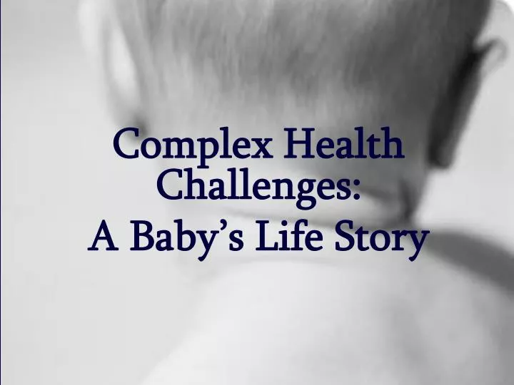
Complex Health Challenges: A Baby’s Life Story. Jessica Potts is a 32 year old woman who is pregnant for the first time. She is working full time but she and her partner, Susan, are just making ends meet with the expenses for IVF. .

E N D
Complex Health Challenges: A Baby’s Life Story
Jessica Potts is a 32 year old woman who is pregnant for the first time. She is working full time but she and her partner, Susan, are just making ends meet with the expenses for IVF.
Jessica is an average height and weight woman as is Susan. When they decided to have a child it was discovered that Susan has endometriosis and fibroids and can not get pregnant therefore Jessica will be carrying the child.
Treatment of infertility • Usually does not fully prevent conception, especially in mild to moderate cases • Infertility is more common in women with severe forms of disease • Treatments are varied • Surgical treatments are superior to hormonal or medical treatments when goal is enhanced fertility • Assisted reproduction may be used
After being unsuccessful with artificial insemination, the couple opt for in vitro fertilization, Jessica is successful with IVF therapy on the first attempt.
IVF In vitro fertilization Successfully used in 1978
IVF: Who May Benefit? • Women who have a blocked or damaged fallopian tube • Mild problem with male partner’s sperm • No cause identified for inability to conceive • Patient’s who have tried IUI or ovulation induction with no success
IVFProcedure • Fertility drugs: -gonadotrophin-releasing hormones (GnRH) -human-menopausal gonadtrophin (hMG) -human chorionic gonadotrophin (hCG). • Monitor blood hormone levels • Ultrasound scan • Remove ova through ultrasound-guided transvaginal retrieval or laparoscopy
IVF Procedure Cont’d Eggs are mixed with sperm in dish and cultured in incubator Dish checked in 2 days to see if eggs have been fertilized Those kept are kept for a couple more days and checked again Fertilized eggs form ball of cells-embryo Healthiest embryo is inserted into uterus Taking progesterone all along to thicken lining
IVF Procedure Cont’d Progesterone given by injection, pessary (gel) Endometrium too thin IVF cycle abandoned Two embryos transferred with thin catheter through cervix into the uterus (via ultrasound) No more then three embryos can be legally transferred Number of embryos transferred depends on your age and chances of success If successful able to take a pregnancy test in 2 weeks
IVF • IVF treatment takes 4-6 weeks to complete • Success rates vary • Advantages: • gives women with blocked, damaged, or missing fallopian tubes a chance to have a baby • Disadvantages: • increased chances for multiple births, increase risk for miscarriage and other complications, hormones not closely monitored lead to ovarian hyperstimulation syndrome, ectopic pregnancy
MULTIPLE PREGNANCY Multiply pregnancy occurs when the use of ovulation inducing medication triggers the release of multiple eggs, which, when fertilized produce multiple embryos that are then implanted
Case Study • The couple live in a small town about half an hour from the city. • The medical clinic arranges for Jessica’s prenatal care and any tests and assessments she may need for the duration of her pregnancy and delivery.
Case Study • Jessica and Susan are so happy to finally be pregnant, they refuse most prenatal tests. • They’re just so happy to have a baby in their lives, they are not too concerned about genetics and anomalies with the baby. • They due, however, agree to have an ultrasound.
Doppler Ultrasound This noninvasive test measures blood flow in different parts of your baby's body — such as the umbilical cord, brain, liver, and heart — to help your caregiver assess your baby's health. It can be done at the same time as an ultrasound and uses the same equipment. Student presentation for more information…
Case Study Mrs. Potts goes into spontaneous labor at 3am at 39 weeks gestation. Her labor and delivery are unremarkable and Jessica and Susan welcome baby Isabelle at 5:56am. The couple become concerned when the nurses start whispering about the baby. Jessica keeps asking them what the problem is, but gets no immediate response. One nurse finally tells Susan and Jessica that the doctor will be in soon to talk to them. Eventually, the doctor delivers the news that their precious little baby girl infact has Down Syndrome. Jessica and Susan react with anger. They are dissappointed that they didn’t go through with the serum screening tests however, they must move forward now.
Down Syndrome … • Is the most frequently occurring chromosomal disorder • Is universal across race and gender • Is caused by an error in cell division • Occurs at conception • Why it occurs is unknown
History • John Langdon Down (the father of down syndrome) • 1866: published an accurate description of a person with down syndrome • Jerome Lejeune • Identified down syndrome as a chromosomal anomaly
Incidence • Incidence increases with age • In Canada Down Syndrome occurs in approximately 1 in 800 live births • Increases to 1 in 100 in second birth if your first child had Down Syndrome • In the US more than 350 000 people have Down Syndrome
Trisomony 21 • Also called Down’s Syndrome • Specific characteristics • Chromosome abnormality • Genetic testing
Down Syndrome • The presence of 47 chromosomes instead of 46 • More specifically it is the presence of extra genetic material associated with the 21st chromosome. (Trisomy) • Caused by an error in cell division
Mitosis • The process of cell division involved in all cell growth, differentiation, and repair • The chromosomes of each cell duplicate • Two daughter cells are produced • They are diploid (contain 46 chromosomes in 23 pairs) • Occurs in all cells except for the oocytes and sperm
Meiosis • Occurs in reproductive cells • There is a reduction in the number of chromosomes occurs (end up with 23 chromosomes) • Oocytes and sperm are referred to as being haploid (contain a single copy of each chromosome) • The paired chromosomes come together in preparation for cell division, portions cross over and genetic material is exchanged • recombination creates greater diversity in oocytes and sperm
Nondisjunction • Accounts for approximately 95% of down syndrome cases • A pair of chromosomes may fail to separate completely creating a sperm or oocyte that contains either 2 copies or no copies of a particular chromosome. • Trisomy: when there are 2 copies • Monosomy: when there are no copies • Prior to or at conception, a pair of 21st chromosomes in either the sperm or the egg fails to separate. As the embryo develops, the extra chromosome is replicated in every cell of the body
Trisomy • Down syndrome is a form of trisomy • there is extra genetic material on the 21st chromosome • Trisomy can occur on any chromosome but the only forms that are frequently seen in live births are on the 13, 18, and 21 chromosome
Mosaicism • Mosaicism occurs when nondisjunction of chromosome 21 takes place in one of the initial cell divisions after fertilization causing a person to have 46 chromosomes in some of their cells and 47 in others • This is the least common form of Down syndrome • accounts for only 1 to 2 percent of all cases
Translocation • Occurs when part of chromosome 21 breaks off during cell division and attaches to another chromosome, usually chromosome 14. While the total number of chromosomes in the cells remains 46, the presence of an extra part of chromosome 21 causes the characteristics of Down syndrome • Maternal age is not linked to the chance of having a baby with translocation. Most cases are sporadic, chance events, but in about one third of translocation cases, one parent is a carrier of a translocated chromosome. For this reason, the chance of translocation in a second pregnancy is higher than that seen in nondisjunction. • Accounts for 3 to 4 percent
Appearance • low nasal bridge • epicanthal folds (eyes) • protruding tongue • low set ears • poor muscle tone (hypotonia) • short stature • single crease across palm of hand • slightly flattened facial profile
Characteristics & Conditions • 3/4 fetuses are spontaneously aborted • 20% die before the age of 10 r/t complications • have and IQ ranging from 25-50 • 1-3 to1-2 have congenital heart defects • most common are an atrioventricular septal defect, persistant ductus arteriosus, and tetraology of fallot • decreased ability to fight respiratory infections • increased susceptibility to leukemia • usually develop Alzheimer symptoms by the age of 40 • increased risk for thyroid and vision problems • usually accompanied by some level of mental retardation • average life expectancy is 55
Treatment • Down syndrome is not treated • The symptoms are treated • It is important to encourage individuals with down syndrome to develop their gifts and talents • Early intervention programs can be initiated • Many individuals with down syndrome go to school (elementary, secondary, and post-secondary) and some adults are capable of working in the community. • With proper care individuals with down syndrome can lead healthy lives
Diagnosis • Prenatal screening (usually diagnosed here, but not in Jessica’s case) • maternal serum screening • ultrasound (sonogram) screening • Diagnostic testing • chorionic villus sampling (CVS) • amniocentesis • percutaneous umbilical blood sampling (PUBS) • Diagnosis is made at the birth of baby Isabelle
Along with the diagnosis of Down’s • The doctor informs Susan and Jessica that he would like to run some tests on baby Isabelle as it appears her head is abnormally large • Jessica and Susan are panicked at this point and the nurse tries to comfort the couples’ anxiety • The doctor refers to a neurologist who examines baby Isabelle
The neurologist is a stoic man in his late 60’s and doesn’t believe in same-sex couples having children • He bluntly tells Susan and Jessica the his diagnosis of baby Isabelle and refers them to community services for follow up • Isabelle has…
What is the Diagnosis? Hydrocephalus
Ventricles of the Brain A Lateral View An Anterior View
Ventricles of the Brain • Lateral Ventricles • Each cerebral hemisphere contains a large lateral ventricle: Right and Left ventricle or First and Second ventricle • The septum pellucidum separates the two lateral ventricles • Third Ventricle • Located in the diencephalons • Two lateral ventricles are not directly connected to each other • communicates with the third ventricle through an interventricular foramen (foramen of Monro) • Third and Fourth ventricle are connected through a slender canal known as the mesencephalic aqueduct (the aqueduct of Sylvius) located in the mesencephalon • Fourth Ventricle • Superior portion lies between the posterior surface of the pons and the anterior surface of the cerebellum • Extends into the superior portion of the medulla oblongata • Then narrows and becomes continuous with the central canal of the spinal cord
Ependymal Cells • Central canal: narrow passageway in the spinal cord • In the brain, the passageway forms the ventricles • The central canal and ventricles are lined by a cellular layer of epithelial cells called the ependyma and are filled with cerebrospinal fluid (CSF) • During embroyonic development and early childhood, the free surface of ependymal cells are covered with cilia • The cilia persists in adults only within the ventricles of the brain, where they assist in the circulation of CSF • In other areas, the ependymal cells typically have scattered microvilli • Function: • participate in the secretion of the CSF • sensory functions, such as monitoring the composition of the CSF
The Cranial Meninges Layers: • Cranial dura mater: • Consists of outer (Endosteal) and inner (Meningeal) fibrous layers • Layers are typically separated by a slender gab that contains tissues fluids and blood vessels, including several large venous sinuses (Dural sinus). • The veins of the brain open into these sinuses, which deliver the venous blood to the internal jugular veins in the neck. • Arachnoid: • Consists of the arachnoid membrane, an epithelial layer, and the cells and fibers of the arachnoid trabeculae that cross the subarachnoid space to the pia mater. • Arachnoid membrane covers the brain, providing a smooth surface that does not follow the brain’s underlying folds. • Pia mater: • Sticks to the surface of the brain • It extends into every fold, and accompanies the branches of cerebral blood vessels as they penetrate the surface of the brain to reach internal structures.
The Cranial Meninges Function: • To protect the brain • Dural folds provide additional stabilization and support to the brain. • Dural sinuses are large collecting veins • Three layers of dural folds: • Falx cerebri: • Superior sagittal sinus and the inferior sagittal sinus (venous sinuses) lie within this dural fold. • Tentorium cerebelli: • Transverse sinus lies within the tentorium cerebelli. • Falx cerebelli
Cerebrospinal Fluid Function: • Completely surrounds and bathes the exposed surfaces of the CNS and has several important functions: • Cushioning Delicate Neural Structures • Supporting the Brain: • The brain is suspended inside the cranium and floats in the CSF. • A human brain weighs about 1400 g in the air, but only about 50 g when supported by the CSF. • Transporting Nutrients, Chemical Messengers, and Waste Products: • Ependymal lining is freely permeable (exception: choroids plexus) • CSF is in constant chemical communication with the interstitial fluid of the CNS