Download

1 / 32
650 likes | 2.27k Views
Antigen-antibody reaction. Antigen = immunogen : any foreign substance which, when introduced will evoke a specific immune response. In terms of infectious diseases, the following may act as antigens:

E N D
Antigen= immunogen: any foreign substance which, when introduced will evoke a specific immune response. In terms of infectious diseases, the following may act as antigens: • Microbial structures (cell walls, capsules, flagella, pili, viral capsids, envelope-associated glycoproteins, etc.). • Microbial exotoxins. • Certain noninfectious materials may also act as antigens if they are recognized as "nonself" by the body. Theseinclude: • Allergens. • Foreign tissues and cells. • The body's own cells that the body fails to recognize as "normal self" as cancer cells.
Antibodies or immunoglobulinsare specific protein configurations produced by B-lymphocytes and plasma cells in response to a specific antigen and capable of reacting with that antigen. Antibody structure (IgG)
Antigen-antibody reactions can be used in the laboratory to identify unknown microorganisms
Antigen antibodies reaction • agglutination • precipitation • complement fixation • Toxin antitoxin neutralization • virus neutralization • Immunefluorescence • Flow cytometry • Enzyme linked immunesorbant assay (ELISA) • Radioimmunoassay
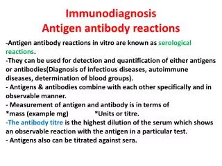
Serologic testing may be used in the clinical laboratory in two distinct ways: • To identify unknown antigens (such as microorganisms). • To detect antibodies being made against a specific antigen in the patient's serum.
Agglutination • Antigen: suspension of microorganisms, cells or latex particles coated with antigens. • Antibodies: specific anti serum • Reaction …………………… Agglutination or clumping. If one (antigen or antibody) is known, the reaction can be used for identification of the other.
I. Direct agglutination 1. Slide agglutination: • Antigen + Antibody Clumping occurs if the serum is specific to the organism. Blood grouping
2. Tube agglutination • It is a quantitative test. • Used to determine the amount of antibodies in the patient’s serum. titre is Serial dilution of the patient serum showing agglutination
Widal test Serological method used for diagnosis of enteric fever. The idea is to detect the antibodies to salmonella in patient’s serum. This test demonstrates the presence of somatic (O) and flagellar (H) agglutinins to Salmonella typhiin the patient's serum using suspensions of O and H antigens. Antigens of S. paratyphi A, S. paratyphi B, S. paratyphi C are included in most commercial kits. We can do by using : • Slide agglutination method. • Tube agglutination method.
Slide method: Slide Widal test is more popular among diagnostic laboratories as it gives rapid results. I. Qualitative test: • Dispense 0.08 of undiluted patient serum sample onto a raw of circles. • Add one drop of each O or H antigen suspension to the serum (shake the reagent bottle before use) . • Mix well using a stirring stick gently rotate the slide for one minute. • Appearance of agglutination gives qualitative positive results.
II. Quantitative test (slide titration method) : • 80 μl, 40 μl, 20 μl, 10 μl and 5 μl of patient’s serum for each antigens are placed on the slides. • To each series of serum specimen, one drop of specific antigen is added. • Mixed well and rotate the slide for one minute. • Agglutination in each of these is noted. 80 μl corresponds to 1in 20 dilution, 40 μl to 1 in 40, 20 μl to 1 in 80, 10 μl to 1 in 160 and 5 μl corresponds to 1 in 320 titre.
Tube agglutination method Prepare 8 sets each is prepared as follows: • 8 plastic tubes in a rack. • 1.9 ml of saline in the 1st tube and 1 ml into the remaining seven. • Dispense 0.1 ml of the of the serum in 1 st tube and mix well. • Dispense 1 ml from 1st tube into 2nd tube and mix well. • Continue this doubling of dilutions to the other tubes. • Discard 1 ml from the last tube. • Shake the reagent bottle well. • Add one drop of the antigen suspension into each tube and mix. • Incubate .
10. Examine for agglutination. 11. The titre to be taken is the highest dilution tube to show agglutination.
Titres below 1/80 are of no significance (indicates previous sub clinical infection) due to the endemicity of the disease in the area. • Agglutination of O suspension indicates recent infection. • Recently vaccinated individuals will possess agglutinins to S. typhi and paratyphi. The agglutination will occur with more than one suspension. • For proper interpretations of the widal test, two serum samples separated by ten days should be tested and the rising titre in the second serum sample is indicative of active enteric fever.
II.Passive (indirect) agglutination What is a difference between passive and active agglutination? • In active agglutination you have a particulate Ag + Ab, since the Ag is particulate, large, when a complex is formed it is visible. • In passive agglutination the Ag is soluble so it must first be attached to something like latex beads, red blood cells so when agglutination occurs it can be seen with the naked eye.
Passive agglutiniation include: • Pregnancy test. • ASO testing. • C- reactive protein testing. • Rapid plasma reagen test. • Infectious mononucleosis latex agglutination test. • H. pylori testing.
1. Latex method pregnancy test • It depends on the appearance of HCG in blood & urine of pregnant females. • Method: Drop of latex particles coated with anti HCG + drop of urine Agglutination
2. ASO ( anti streptolysin O) testing • Slide agglutination test performed for patients with streptococcal ß hemolytic infections. • Latex particles coated with SO antigens + serum containing Abs Agglutination
3. CRP (C- reactive protien) • Slide agglutination test. • Latex particles coated with specific antihuman C- reactive protein antibodies + serum containing CRP. Agglutination
4. RPR (Rapid plasma reagen) • Slide test used for diagnosis of sypillis. • Syphillis is a venereal disease caused by Treponemapallidum. • The test measures antibodies against the lipoidal material released from the damaged host cell, so it is not very specific. • Specific Ag containing micro particulate carbon + Serum containig the Abs Agglutination
5. Infectious mononucleosis latex agglutination (IM Latex Test) • Latex agglutination slide test used for determination of infectious mononucleosis in non-diluted serum or plasma. • Infectious mononucleosis is a disease caused by the Epstein Barr virus.
The IM latex test provides: • Infections with IM is associated with production of heterophile antibodies. • These heterophile antibodies can cross react with glycoprotien on the surface of the sheep or bovine RBCS . • Latex particles coated with partially purified glycoprotein (from bovine red blood cells). + Serum which contains heterophile antibodies associated with IM Agglutination.
Agglutination identifies the presence of the heterophile antibodies in the sample (It is a non specific test that can be used for screening of patients).
6. One step H. pylori Test • H. pylori is a small, spiral-shaped bacterium that lives in the surface of the stomach and duodenum. • This test is a rapid one step test for the qualitative detection of antibodies to Helicobacter pylori (H. pylori) in human serum or plasma. • The one step H.pylori Test device is a simple test that utilize a combination of H.pylori antigen coated particles and anti-human IgG to detect H.pylori antibodies in serum or plasma.
In this test procedure: • Anti human IgGis immobilized in the line region of the test. Specimen contain H.pylori antibodies will be added to the “specimen well” of the device, it will react with H.pylori antigen coated particles in the test. • This mixture migrates chromatographically along the length of the test device and interacts with the immobilized anti-human IgG. • If the specimen contains H.pylori antibodies, a colored line will appear in the test line region indicating a positive result.
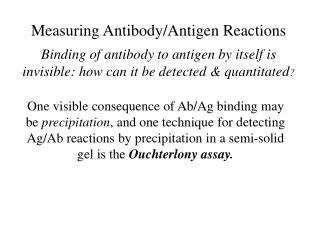
Precipitation • This is an Ag-Ab reaction in which the Ag is soluble (bacterial toxin). • When antigens and antibody mixed in the proper proportion, they form large macromolecular complexes called precipitates.
Tube precipitation Used in estimation of C3. CRP, immunoglobulins.
Agar gel diffusion • Diffusion of Ag & Ab is allowed to occur in agar gel. • Double diffusion Ag & Abs are put in wells in the agar. • Used in grouping of Streptococci ( groups: A, B, C, D). • Eleck’s test: fordiphtheria Strain toxin production. • Single diffusion Ab is mixed with the agar before pouring in plates and the Ag is placed in agar wells Precipitation lines