Download
1 / 41
410 likes | 624 Views
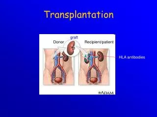
Transplantation. Transplantation = transfer of tissue or organ ● autologous - donor = recipient ● syngeneic - genetically identical donor and recipient (identical twins) ● allogeneic - genetically nonidentical donor of the same species

E N D
Transplantation = transfer of tissue or organ ●autologous - donor = recipient ● syngeneic - genetically identical donor and recipient (identical twins) ● allogeneic - genetically nonidentical donor of the same species ● xenogenic - the donor of another species ● implant - artificial tissue compensation
Allotransplantation ● differences in donor-recipient MHC gp and secondary histocompatibility Ag ●alloreactivity of T lymphocytes - the risk of rejection and graft-versus-host disease ●direct detection of alloantigens – recipient T lymphocytes recognize the different MHC gp and non-MHC molecules on donor cells ●indirect recognition of alloantigens - APC absorb different MHC gp from donor cells and present the fragments to T lymphocytes ● CD8+ T cells recognize MHC gp I. ● CD4+ T cells recognize MHC gp II.
Testing before transplantation ●ABOcompatibility -risk of hyperacute or accelerated rejection (= formation of Ab against A or B Ag on graft vascular endothelium) ●HLA typing (determining of MHC gp alelic forms) phenotyping and genotyping by PCR ●Cross-match - lymphocytotoxic test – detection of preformed Ab (after blood transfusions, transplantation, repeated childbirth) ●Mixed lymphocyte reaction - testing of T lymphocytesalloreactivity, monitor for reactivity of lymphocytes to allogeneic HLA
HLA typing = determmination of HLA antigens on the surface of lymphocytesCarry out during the testing before transplantation and in determination of paternity • 1) Serotyping • Microlymfocytotoxic test • Allospecific serums (obtained from multiple natal to 6 weeks after birth, obtained by vaccination of volunteers, or commercially prepared sets of typing serums (monoclonal antibodies)) • Principle - the incubation of lymphocytes with typing serums in the presence of rabbit complement, then is added the vital dye which stained dead cells - cells carrying specific HLA are killed by cytotoxic Ab against the Ag, the percentage of dead cells is a measure of serum toxicity (forces and antileukocyte antibody titre) • Positive reaction is considered more than 10% dead cells (serological typing can be done also by flow cytometry
2) Molecular genetic methods • For typing are used hypervariable sections in the II. exon genes coding for HLA class II; to determine HLA class I is used polymorphism in II. and III. exon coding genes2a) PCR-SSP= Polymerase chain reaction with sequential specific primers • Extracted DNA is used as a substrate in a set of PCR reactions • Each PCR reaction contains primers pair specific for a certain allele (or group of alleles) • Positive and negative reactions are evaluated by electrophoresis, each combination of alleles has a specific electrophoretic painting
2b) PCR-SSO • PCR reaction with sequence-specific oligonucleotides Multiplication of hypervariable sections of genes coding HLA • Hybridization with enzyme or radiolabeled DNA probes specific for individual alleles 2c) PCR-SBT • Sequencing based typing • The most accurate method of HLA typing • We get the exact sequence of nucleotides, which compares with a database of known sequences of HLA alleles
Cross-match testing ● determination of preformed antibodies ● recipient serum + donor lymphocytes + rabbit complement → if cytotoxic Ab against donor HLA Ag are present in recipient serum (called alloantibodies = Ab activating complement) → lysis of donor lymphocytes. Visualization of dye penetration into lysis cells. ●positive test = the presence of preformed Ab → risk of hyperacute rejection! → contraindication to transplantation
Mixed lymphocyte reaction (MRL) ● determination of T lymphocytesalloreactivity ● mixed donor and recipient lymphocytes → T lymphocytes after recognition of allogeneic MHC gp activate and proliferate One-way MRL ● determination of recipient T lymphocytesreactivity against donor cells ● donor cells treated with chemotherapy or irradiated lose the ability of proliferation
Immunologically privileged sites and tissues • Transplantation of some tissues don´t lead to the induction of allogeneic reactivity • minimal content of lecocytes • mechanisms that prevent to the development of injurious inflammation • Evolutionarily significant, protection of vital organs (brain, eye, gonads) • Factors protecting immunologically privileged structures • isolation from the immune system • preference of TH2 reactoin, supression of TH1 reaction • FasL expression • production of TGFb
Hyperacute rejection ● minutes to hours after transplantation ●humoral mediated immune response mechanism: ● if in recipients blood are present preformed or natural Ab (IgM anti-carbohydrate Ag) before transplantation→ Ab + Ag of graft (MHC gp or endothelial Ag) → graft damage by activated complement (lysis of cells) ● the graft endothelium: activation of coagulation factors and platelets, formation thrombi, accumulation of neutrophil granulocytes prevention: ● negative cross match before transplantation, ABO compatibility
Accelerated rejection ● 3 to 5 days after transplantation ● caused by antibodies that don´t activate complement ●cytotoxic and inflammatory responses triggered by binding of antibodies to Fc-receptors on phagocytes and NK cells prevention: ● negative cross match before transplantation, ABO compatibility
Acute rejection ● days to weeks after the transplantation or after a lack of immunosuppressive treatment ●cell-mediated immune response mechanism: ● reaction of recipient TH1 and TC cells against Ag of graft tissue ● infiltration by lymphocytes, mononuclears, granulocytes around smallvessels → destruction of tissue transplant
Chronic rejection ● from 2 months after transplantation ● the most common cause of graft failure mechanism is not fully understood: ● non-immunological factors (tissue ischemia) and TH2 responses with production alloantibodies, pathogenetic role of cytokines and growth factors (TGF β) ● fibrosis of the internal blood vessels of the transplanted tissue, endothelial damage →impaired perfusion of graft → gradual loss of its function dominating findings: vascular damage
Rejection Factors: ●The genetic difference between donor and recipient, especially in the genes coding for MHC gp (HLA) ●Type of tissue / organ - the strongest reactions against vascularized tissues containing many APC (skin) ●The activity of the recipientimmune system - the immunodeficiency recipient has a smaller rejection reaction; immunosuppressive therapy after transplantation – suppression of rejection ●Status of transplanted organ - the length of ischemia, the method of preservation, traumatization of organ at collection
Bone Marrow Transplantation ● Removal of hematopoietic stem cells● Myeloablation● Transplantation● Engraftment● Rejection● Graft-versus-host reaction
Graft-versus-host (GvH) disease ● after bone marrow transplantation ● GvH also after blood transfusion to immunodeficiency recipients ● T-lymphocytes in the graft bone marrow recognize recipient tissue Ag as foreign (alooreactivity)
Acute GvH disease ● days to weeks after the transplantation of stem cells ● damage of liver, skin and intestinal mucosa ● prevention: appropriate donor selection, the removal of T lymphocytes from the graft and effective immunosuppression
Chonic GvH disease ● months to years after transplantation ● infiltration of tissues and organs by TH2 lymphocytes, production of alloantibodies and cytokines → fibrosis ● process like autoimmune disease: vasculitis, scleroderma, sicca-syndrome ● chronic inflammation of blood vessels, skin, internal organs and glands, which leads to fibrosis, blood circulation disorders and loss of function
Graft versus leukemia effect (GvL) ● donor T lymphocytes react against residual leukemick cells of recipient (setpoint response) ● mechanism is consistent with acute GvH ● associated with a certain degree of GvH (adverse reactions)
Immunologic relationship between mother and allogenic fetus ●fetal cells have on the surface alloantigens inherited from his father ●pregnancy = "semiallogenic transplantation“ Tolerance of fetus by mother allow the following mechanisms: ●the relative isolation of the fetus from maternal immune system (no mixing of blood circulation) ●trophoblast - immune barrier witch protects against mother alloreactive T lymphocytes - don´t express classical MHC gp, expresses non-classical HLA-E and HLA-G ● transfer of small doses of fetal antigens in maternal circulation causes tolerance ... suppressin of TH1 and preference of TH2 immune mechanisms in pregnancy
Rh incompatibility • Complications in pregnancy: production of anti-RhD antibodies by RhD- mother carrying an RhD+ fetus (hemolytic disease of newborns) • Fetal erythrocytes penetrate into the maternal bloodstream during pregnancy - a small amount, don´t immunize • During childbirth or abortion (after 8 weeks of gestation) fetal erythrocytes can penetrate into the bloodstream of mother → immunization, formation of anti-RhD antibodies • After childbirth, investigate Rh factor of born child, if is child Rh+, mother gets up to 72 hours after birth injection of anti-Rh antibodies (administered after abortion too)
Anti-Rh(D) injection, this antibodies bind to RhD Ag on baby´s red blood cells, this Ag than can´t bind to BCR and can´t activate B lymphocytes, this immune comlexes also actively inhibit B lymphocytes • During next childbirths, if fetus is Rh+ and mother produce anti-Rh antibodies, this Abb destroy red blood cells of fetus, which can lead to fetal death, or in severe postpartum anemia(anemia neonatorum) and neonatal jaundice (icterus gravis neonatorum) • For each pregnant woman during the first trimester investigate blod Rh factor and the presence of antibodies, in Rh- women performed a test for antibodies also in II. and III. trimester
Tumor antigens • Antigens specific for tumors (TSA) • complexes of MHCgp I with abnormal fragments of cellular proteins- chemically induced tumors - leukemia with chromosomal translocation • complexes of MHCgp with fragments of proteins of oncogenic viruses- tumors caused by viruses (EBV, SV40, polyomavirus) • abnormal forms of glycoproteins- sialylation of surface proteins of tumor cells • idiotypes of myeloma and lymphoma- clonotyping TCR and BCR
b) Antigens associated with tumors (TAA) • present also on normal cells • differences in quantity, time and local expression • auxiliary diagnostic markers 1)onkofetal antigens • on normal embryonic cells and some tumor cells • -fetoprotein (AFP) - hepatom • carcinoembryonic antigen (CEA) - colon cancer 2) melanoma antigens • MAGE-1, Melan-A
3) antigen HER2/neu • receptor for epithelial growth factor • mammary carcinoma 4) EPCAM • epithelial adhesion molecule • metastases 5) differentiation antigens of leukemic cells • present on normal cells of leukocytes linage • CALLA -acute lymphoblastic leukemia (CD10 pre-B cells)
Anti-tumor immune mechanisms Immune control • tumor cells normally arise in tissues and are eliminated by T lymphocytes • probably wrong hypothesis Defensive immune response • tumor cells are weakly immunogenic • occurs when tumor antigens are presented to T lymphocytes by dendritic cells activated in the inflammatory environment • if tumor cells are detected, in defense may be involved non-specific mechanisms (neutrophilic granulocytes, macrophages, NK cells) and antigen-specific mechanisms (complement activating antibodies or ADCC, TH1 and TC)
DC are necessary for activation of antigen specific mechanisms • cancer-associated antigens are processed by DC and recognized by T lymphocytes in complex with HLA I. and II. class with providing costimulus signals • predominance of TH1(IFN g, TNFa) • specific cell-mediated cytotoxic reactivity –TC • activation of TH2 → support B lymphocytes→ tumor specific antibodies (involved in the ADCC) • tumor cells are destroyed by cytotoxic NK cells (ADCC) • interferons - antiproliferative, cytotoxic effect on tumor cells - INFγ - DC maturation
Regulatory T cellsprevents removal of cancer cells and thus contribute to the development of the tumor.
Mechanisms of tumor resistance to the immune system - • high variability of tumor cells • low expression of tumor antigens • sialylation • tumor cells signals do not provide costimulus → T lymphocyte anergy • some anticancer substances have a stimulating effect • production of factors inactivating T lymphocytes • expression of FasL → T lymphocyte apoptosis • inhibition of the function or durability dendritic cells (NO, IL-10, TGF-b)
Tumor immunotherapy Therapy- surgical removal of tumor - chemotherapy or radiotherapy - immunotherapy Immunotherapy-induction of anti-tumor immunity, or the use of immune mechanisms to targeting drugs to the tumor site
Immunotherapy using antibodies Antibodies functions - opsonization - activation of complement - induction of ADCC - carriers of drugs or toxins
1) Monoclonal antibodies - against TAA - mouse and humanised antibodies - imunotoxins, radioimunotoxins - the possibility of damage surrounding tissues - HERCEPIN - Ab against HER2/neu, breast cancer - RITUXIMAB - Ab against CD20, lymphoma 2) Bispecific antibodies - bind a tumor antigen and the T lymphocyte or NK cell - Fc fragment of antibody binds to Fc receptors on phagocytes and NK cells 3) Elimination of tumor cells from the suspension of bone marrow cellsusing monoclonal antibodies for autologous transplantation
Immunotherapy using cell-mediated mechanisms 1) stimulation of inflammation at the tumor site 2) stimulation of LAK and TIL - isolation of T and NK cells, stimulation by cytokines, and return to the patient - LAK (lymphokine activated killers) - TIL (tumor infiltrating lymphocytes) 3)improving of tumor cells antigenpresenting function - genetic modification of tumor cells - expression of CD80, CD86 - production of IL-2, GM-CSF - modified cells are irradiated and returned to the patient
4) the dendritic cell immunotherapy- in vitro cultivation of monocytes in an appropriate cytokine environment (GM-CSF, IL-4) → transformation into dendritic cells- cultivation of dendritic cells with tumor antigens
5)tumor vaccines - in vitro stimulation of TH1 cells and TC with tumor antigens 6) immunotherapy by donor T lymphocytes - after allogeneic transplantation - causing graft-versus-host disease 7) immunotherapy by immune system products - IL-2 - renal cell carcinoma • IFN - hematoonkology • 8) Anti CTLA-4 antibody • - Treg inhibition, longer activation of effector T cells