Download

1 / 58
590 likes | 864 Views
Acute upper airway obstruction. Prepared by Ghassan Al-Maimani. Upper Airways. Lower Airways. DEFINITION. Obstruction of the portion of the airways located above the thoracic inlet. EXTENT. Ranges from nasal obstruction till larynx and upper trachea. Clinical manifestation.

E N D
Acute upper airway obstruction Prepared by Ghassan Al-Maimani
Upper Airways Lower Airways
DEFINITION Obstruction of the portion of the airways located above the thoracic inlet. • EXTENT Ranges from nasal obstruction till larynx and upper trachea.
Clinical manifestation • Stridor : ( Inspiratorystridor ) - Harsh sound produced by vibration of upper airway structure - Indicates upper airway obstruction • Hoarseness: Indicates involvement of vocal cords • Respiratory distress / suprasternal retraction
Clinical manifestation : cont. • Cough • Signs of hypoxemia -Anxiety - Restlessness - Tachycardia - Pallor - Cyanosis: late sign
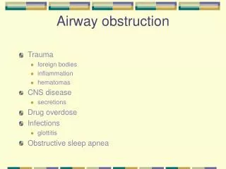
Causes of acute UAO : • Infectious • Non- Infectious ( commonest )
INFECTIOUS • Croup ( Acute laryngotracheobronchitis ). • Bacterial trachitis( membranous croup ). • Acute epiglottitis. • Diphtheria. • Retropharyngeal abscess / peritonsillar. abscess.
Non-INFECTIOUS • Foreign body inhalation. • Spasmodic laryngitis • Caustic burn and trauma.
Croup ( laryngotracheobronchitis) Term applied to group of inflammatory conditions involving larynx , trachea and characterized by Triad : • Inspiratory stridor • Brassy cough • Hoarseness of voice +/_ resp.distress
Usually viral in origin - Parainfluenza virus (type 1) - Influenza virus - RSV , adenovirus , measles virus • It is the most common cause of Acute Airway Obstruction in children • Age group 3m-3 years (peak 2years) • Affects boys more often than girls • Peak occurrence is in fall and winter
Clinical features • Usually h/o preceding URTI • Gradual or sudden in onset • Triad : Inspiratory stridor Brassy cough Hoarseness of voice +/_ resp.distress
Diagnosis • It is clinically diagnosed • Neck x-ray and CBC all should be done in clinically stable pt . - AP neck film : show a pencil tip or steeple sign of the subglottic trachea - CBC , it may helps .
Do not use a radiograph to make management decisions in a pt. with an unstable airway
Treatment - Some children improve spontaneously because of natural fluctuations in the disease - Mist therapy / Steam inhalation Oxygen Adequate hydration Nebulization with Racemic epinephrine
Steroid • Used in moderate to severe croup • A child who needs admission in ICU for croup management needs steroid. • Preparations • Dexamethasone • Nebulized Budesonide • Not as effective as dexamethasone • Much more expensive than dexamethasone
Do we use steroid in mild croup ? for Children with mild croup , dexamethasone is an effective treatment that results in consistent and small but important clinical and economic benefits ( level Ib)
Which is more effective oral or nebulized dexamethasone for children with mild croup ? Children with mild croup who receive oral dexamethasone Rx are less likely to seek subsequent medical care and demonstrate more rapid symptom resolution compared with children who receive nebulized dexamethasone or placebo Rx ( level Ib )
Most children with croup doesn't need hospitalization because symptoms typically resolve within a few days
ICU admission • Signs of hypoxia • Severe distress with exhaustion • Decision about ventilation
Bacterial causes of acute airway obstruction • Acute epiglottitis --- Hemophilusinfluenzae type B • Bacterial tracheitis --- Staph Aureus • Cornybactrium diphtheria
Acute epiglottitis • It is a rapidly progreesive bacterial infection causing acute inflammation and edema of the epiglottis and adjacent structures : aryepiglottic folds and arytenoids • Also known as supraglottitis • It is life threatening condition may lead to sudden and complete airway obstruction
Age : 2-6 years ( peak at 3 year) • Infant , older children and adult are rarely affected • Causative agents : - HIB - pneumococci , staphylococci, streptococci
Clinical features • Previously well child • Sudden onset , history is short, 4-12 hours of sore throat and high fever • 4 “ D” Distress Dysphagia Dysphonia Drooling of saliva • may lead to death if complete airway obstruction
Diagnosis • History • Presentation • Appearance of the child Pharynx examination at this stage in ER is absolutely contraindicated • Next step = admission in ICU • Neck x-ray : Not the priority Do not leave the patient unattended
Management • Protection of the airways is the primary priority • Quickly proceed with epiglottitis protocol • It is better to initiate a “false” epiglottitis drill than to miss this disease
Epiglottitis protocol - Safe and supervised transfer to skilled hand - Inform consultant Pediatrics, ENT, ICU, Anesthesia - Don't attempt to examine throat in ER - Keep patient as comfortable as possible - Administering 100% O2
Epiglottitis protocol, cont. - Assembling at bedside CPR equipment including resuscitation bag and mask, intubation equipment - Taking the pt. to OR -Attempt IV line or sampling only after intubation in OR /or Tracheostomy
* After epiglottitis protocol has been performed and pt has secure airways you can do : - blood culture : usually positive for HIB - CBC : WBC may be moderately elevated - lateral neck radiograph : shows a thickened epiglottis ( thumb sign )
Diagnosis confirmed by seeing an edematous cherry-red epiglottis on endoscopy • Endoscopic examination should not be performed in advance of the epiglottitis protocol
The main components of Rx is : - maintain adequate airways until inflammation and edema resolve often 36-72hrs - ParentralAbx directed agiants HI assuming this is the cause : ceftriaxone or cefotaxime if not available may use chloramphenicol - Duration of Rx : 7-10 days
Prophylaxis if there is another child in the house ≤ 4 y not vaccinated to HI give Rifampicin to all family members
Bacterial trachitis • It is uncommon infectious cause of acute UAO • pt may present with croup like symptoms • Etiology : Staph Aureus • On intubation: copious thick secretion ( pus) • with appropriate airway support and Abx most pt . Improve within 5 days
Spasmodic laryngitis • Also known as recurrent croup • Presentation like acute onset of croup • No h/o fever or viral infection • Etiology = Allergic in nature • May develop asthma or atopy later on • It typically resolves spont. • rarely associated with severe RD
In a patient with severe airway obstruction Don’t • inspect the oropharynx • send the patient to radiology for a lateral neck or chest X-Ray • insert an IV • take blood gases
Do’s • Be calm and confidant • Transfer the baby to ICU settings • Let the baby be in mother’s lap or beside mother to make him clam and comfortable • Observe the signs of hypoxia or deterioration • In severe cases or respiratory failure: secure the airway ( intubation / trachesotomy)
Foreign body inhalation • Essentials of diagnosis Acute onset of cyanosis and choking *Inability to cough or vocalize (complete obstruction) *Drooling with stridor (partial obstruction) • Risk age group: 6months-4 years of age
Complete obstruction • Unable to speak • Unable to breath • Unable to cough
Treatment • Children should be allowed to use their own cough reflex to extrude the foreign body in case of partial obstruction. • If obstruction increases acute intervention is needed.
First-aid for a choking baby: Infant <1 year of age: According to AAP and AHA * Place the infant face down over rescue arm with head position below the trunk. Five back slaps are delivered rapidly between infant’s scapula with the heel of hand. * If obstruction persists infant should be rolled over and five rapid chest compression should be performed.