Download
1 / 14
140 likes | 372 Views
Aneurysmal Bone Cysts (ABC’s). Dr. Ted Scriven Sept 8, 2008. ABC’s. Classified as a benign boney lesion More specifically, “benign-aggressive” Benign-aggressive = marked bone destruction, soft tissue extension or pathologic fractures. Etiology. Specific translocation @ 17p13

E N D
Aneurysmal Bone Cysts(ABC’s) Dr. Ted Scriven Sept 8, 2008
ABC’s • Classified as a benign boney lesion • More specifically, “benign-aggressive” • Benign-aggressive = marked bone destruction, soft tissue extension or pathologic fractures
Etiology • Specific translocation @ 17p13 • Can arise de novo, or be associated with another primary: • GCT, chondroblastoma, UBC, osteoblastoma, fibrous dysplasia, nonossifying fibroma, chondromyxoid fibroma, osteosarcoma
Etiology • Result from local circulatory abnormality: Increased venous pressure Local hemorrhage Osteolysis More bleeding • Source of bleeding = capilliaries in cyst membrane • Hemorrhage progresses to destructive lesion
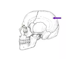
Clinical Picture • Age: often < 20 • Gender: F > M (slight) • Location: • metaphysis or metadiaphysis of long bones (prox humerus, distal femur, prox tibia) • Occasionally iluim or lumbar vertebrae (15 – 20%)
Clinical Picture • Mild pain or swelling • May have neuro deficits with spinal lesions • Duration = weeks years • Symptoms may worsen with pregnancy (more blood volume)
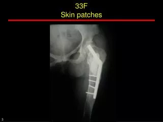
Elevated periosteum Thin shell Investigations • Start with thorough Hx & PE • Xray: • Radiolucent destructive cyst, expands surrounding cortex “Soap-Bubbles” • Often eccentric, can be central or subperiosteal
Investigations • Bone Scan: • Diffuse or peripheral tracer uptake • Central area of decreased uptake • Angiography: • Accumulation of contrast throughout +/- hypervascularity of periphery • Absence of viable afferent or efferent vessels
Investigations • CT • Helps deliniate lesion in areas of complex boney anatomy • MRI • Multiloculated cavities, fluid levels, +/- associated soft tissue mass • Helps to differentiate between ABC & UBC
DDx • UBC • Chondromyxoid Fibroma • Chondroblastoma • GCT • Osteoblastoma • Talengiectatic Osteosarcoma
Pathology • Gross: • Cavitary w/ blood filled spaces • Surrounded by thin layer of bone & raised periosteum
Pathology • Micro: • Hemorrhagic tissue with spaces separated by cellular stroma • No endothelial lining or smooth muscle – only lining is compressed fibroblasts • ALWAYS be sure to examine entire speciman and surrounding area (association with other primaries!!)
Treatment • Curettage & Bone Grafting • Caution: lesion prone to heavy bleeding! • Tourniquet • Pre-op embolization • +/- local adjuvent tx for cavity sterilization: • Phenol, liquid nitrogen, argon • Ressection: • If area is expendable (fibula, metatarsal, etc) • Radiation: • Not routinely used d/t potential for malignant transformation
Prognosis • If primary: • Usually a favourable prognosis • Recurrence: • Rate after curettage = 14 – 34% • Usually within 6/12, rare after 2 yrs • More common in age < 15 yo, centrally located lesions, and when contents not all removed • If associated with another primary: • Classification, treatment and prognosis based on the other (primary) lesion