Download
1 / 35
350 likes | 478 Views
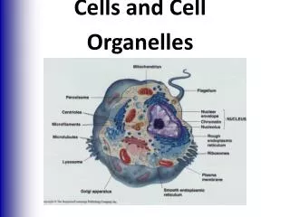
Cells & Tissues. Chapter 3. I. The Plasma Membrane Structure. A. Fluid Mosaic Model. Double bilayer of lipids with imbedded, dispersed proteins Bilayer consists of phospholipids, cholesterol, and glycolipids Glycolipids are lipids with bound carbohydrate

E N D
Cells & Tissues Chapter 3
A. Fluid Mosaic Model • Double bilayer of lipids with imbedded, dispersed proteins • Bilayer consists of phospholipids, cholesterol, and glycolipids • Glycolipids are lipids with bound carbohydrate • Phospholipids have hydrophobic tails and hydrophilic heads
cholesterol: stabilizes lipid membrane • integral proteins: • some face outside of membrane, usually receptors for hormones or other chemical messengers • transmembrane proteins span entire width of membrane & protrude on both sides • transport functions • peripheral proteins: not embedded in lipid • enzymes
glycocalyx: cell coat • stickiness helps bind adjacent cells together • every cell has different pattern of sugars & proteins, biological markers for cell recognition • figure 3.2 p.58
B. Specializations of Plasma Membrane • microvilli: fingerlike extensions of plasma membrane • increase cell membrane surface area • absorption: intestine & kidney
p. 59 figure 3.3 • Tight junctions: binds cell together into leak proof sheets • Intestines prevent digestive enzymes from seeping into bloodstream • Desmosomes: anchoring junctions for cells subjected to mechanical stress • Gap junctions: allow communication between cells
Desmosomes http://www.answers.com/topic/desmosome
A. Membrane Transport • cells are continuously bathed in extracellular fluid called interstitial fluid largely composed of water • contains nutrients, hormones, salts, & waste products
to stay healthy each cell must extract exact amounts of substances needed from this fluid & reject the rest • membrane is selectively permeable: it allows some to pass while excluding others
B. Passive Processes • substances penetrate membrane without any energy input from cell 1. simple diffusion 2. osmosis 3. facilitated diffusion 4. filtration
C. Active Transport Processes • Primary active transport – hydrolysis of ATP phosphorylates the transport protein causing conformational change • Secondary active transport – use of an exchange pump (such as the Na+-K+ pump) indirectly to drive the transport of other solutes
substances are too large or have to move against the concentration gradient 1. Solute Pumping 2. Bulk Transport • endocytosis: phagocytosis or pinocytosis • exocytosis
D. Membrane Potential • Voltage across a membrane • Resting membrane potential (RMP) – the point where K+ potential is balanced by the membrane potential • Ranges from –20 to –200 mV (depends on cell) • Results from differential permeability of the plasma membrane to Na+ and K+ • Steady state – potential maintained by active transport of ions
E. Cell Signaling • Contact signaling – important in normal development and immunity • Electrical signaling – voltage-regulated “ion gates” in nerve and muscle tissue • Chemical signaling – neurotransmitters bind to chemically gated channel-linked receptors in nerve and muscle tissue • G protein-linked receptors – ligands bind to a receptor which activates a G protein, causing the release of a second messenger, such as cyclic AMP or Tyrosine Kinase.
Amino Acid-Based Hormone Action: cAMP Second Messenger Extracellular fluid Hormone A Adenylate cyclase Receptor Gs Catecholamines ACTH FSH LH Glucagon PTH TSH Calcitonin Cytoplasm Figure 16.2
Amino Acid-Based Hormone Action: cAMP Second Messenger Extracellular fluid Hormone A Adenylate cyclase 1 Receptor Gs Catecholamines ACTH FSH LH Glucagon PTH TSH Calcitonin Cytoplasm Figure 16.2
Amino Acid-Based Hormone Action: cAMP Second Messenger Extracellular fluid Hormone A Adenylate cyclase 1 2 GTP Receptor Gs GDP GTP Catecholamines ACTH FSH LH Glucagon PTH TSH Calcitonin Cytoplasm Figure 16.2
Amino Acid-Based Hormone Action: cAMP Second Messenger Extracellular fluid Hormone A Adenylate cyclase 1 2 3 GTP GTP Receptor Gs GDP GTP Catecholamines ACTH FSH LH Glucagon PTH TSH Calcitonin Cytoplasm Figure 16.2
Amino Acid-Based Hormone Action: cAMP Second Messenger Extracellular fluid Hormone A Adenylate cyclase 1 2 3 GTP GTP 4 Receptor Gs GDP GTP ATP cAMP Catecholamines ACTH FSH LH Glucagon PTH TSH Calcitonin Cytoplasm Figure 16.2
Amino Acid-Based Hormone Action: cAMP Second Messenger Extracellular fluid Hormone A Adenylate cyclase 1 2 3 GTP GTP 4 Receptor Gs GDP GTP ATP cAMP Catecholamines ACTH FSH LH Glucagon PTH TSH Calcitonin 5 Triggers responses of target cell (activates enzymes, stimulates cellular secretion, opens ion channels, etc.) Inactive protein kinase A Active protein kinase A Cytoplasm Figure 16.2
III. Body Tissues • Histology: study of tissues • groups of similar cells associated to perform a related function
TYPE 1. Epithelial 2. Connective 3. Muscle 4. Nervous PURPOSE Covering Support Movement Control
IV. EPITHELIAL TISSUES • sheet of cells that covers a body surface or lines a body cavity • cells fit so close to make continuous sheets • contains nerve but no blood vessels • quick regeneration
A. Classification • 1. Simple: single layer of cells, used for absorption or filtration • Squamous: lymph system & lines organs • Cuboidal: exocrine glands and kidneys • Columnar: digestive system