Download
1 / 34
340 likes | 513 Views
Binding Studies on Trafficking Proteins Using Microcalorimetry McMahon lab Neurobiology Division Laboratory of Molecular Biology Cambridge. Binding. Recruitment. Coating. Budding. Uncoating. Clathrin Mediated Endocytosis. Receptor. Ligand. AP adaptor complex. Regulatory adaptor.

E N D
Binding Studies on Trafficking Proteins Using Microcalorimetry McMahon lab Neurobiology Division Laboratory of Molecular Biology Cambridge
Binding Recruitment Coating Budding Uncoating Clathrin Mediated Endocytosis Receptor Ligand AP adaptor complex Regulatory adaptor Clathrin Dynamin
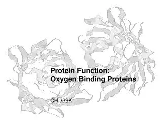
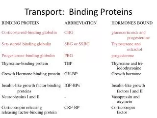
Receptor Mediated Endocytosis a b c Replica of the inner membrane surface (Heuser and Anderson 1989) a) Yolk protein (Gilbert und Perry 1979) b) Low Density Lipoprotein (Anderson et al. 1977) c) Virus particle (Matlin et al. 1981)
, , , 1-4 1-4 1-4 Collins et al. 2002 AP Adaptor Complex Appendage binds regulators Hinge binds clathrin Trunk binds lipids and membrane proteins AP-1 (): TGN / Endosome AP-2 (): Plasma membrane AP-3 (): Lysosome AP-4 (): TGN
AP Trafficking Pathways Plasma membrane AP-2 Endosome Lysosome AP-3 Lysosome-related Organelle AP-4 GGA AP-1 AP-3 Trans-Golgi-Network
AP Appendage Domains DP(F/W) FxDxF DP(F/W) DPW FxxF -Adaptin -Adaptin -Adaptin Owen et al. 2000 Owen et al. 1999 Brett et al. 2002 Kent et al. 2002 Nogi et al. 2002
Eps15 Aminoacid 0 250 500 750 1000 Regulatory Adaptors Epsin1 EpsinR AP180 Dab2 Amphiphysin1 DOMAINPARTNER SH3 PxxPxR PTB Receptor and Lipids ANTH/ENTH Lipids BAR Lipids EH NPF Clathin-Box Clathrin DxF/W - and -Adaptin NPF EH PxxPxR SH3
Interactions in Trafficking Amphiphysin Receptor Lipids FEDNF Yxx or LL AP-Complex LLDLD DxF or FxxF LLDLD DxF or FxxF NPF Epsin1 EpsinR AP180 Dab2 Eps15 Clathrin LLDLD
Determination of Binding Constants Definition of Association and Dissociation Constants: k1 [P]free = conc. of free protein For a binding reaction at equilibrium: P + L PL [L]free = conc. of free ligand k-1 [PL] = conc. of PA complex k1 = rate constant for formation of [PL] k-1 = rate constant for breakdown of [PL] The rate of formation of [PL] is k1 [P]free [L]free, where k1 is a second order rate constant with units of l/mol-1s-1. The rate of breakdown of [PL] is k-1 [PL], where k-1 is a first order rate constant with units of s-1. At equilibrium, the rate of formation of [PL] equals the rate of its breakdown, so k1 [P]free[L]free= k-1 [PL]. Also recall that: KD = k-1 / k1 = [P]free [L]free/ [PL] = 1 / KA KD is given in units of concentration (e.g., mol/l) Or, in terms of fraction of protein binding sites occupied (y), which is often convenient to measure: y = [PL] / ([P]free+ [PL]) • Use [PL] = KA [P]free [L]free •Divide through by KA • Replace KA by 1 / KD = [L]free / ([L]free + KD)
Determination of Binding Constants • Special cases: • y = [L]free / ([L]free + KD) • For [L]free = 0: y = 0 nothing bound • For [L]free: y = 1 full occupancy • For [L]free = KD: y = 0.5 half occupancy • Two possible ways to determine binding constants: • Measure bound and free ligand at equilibrium as a function of concentration • Measure association and dissociation rate constants and use these to calculate binding constants
Methods to determine Binding Constants Signal Information Advantage Disadvantage Spectroscopy change of absorptionKD (10-4-10-11M) in solution probe needed (Fluorescence, UV/Vis, CD) or emission of light Microcalorimetry heat of binding KD (10-3-10-11M) no labels, large sample H, S, nin solution direct access to H direct access to n Surface Plasmon Resonance change ofrefractiveKD (10-3-10-13M) small sample, surface coupled, index due to mass k1, k-1 automated ligand must have large mass Stopped-Flow coupled to spectroscopy KD (10-3-10-12M) fast probe needed k1, k-1 Analytical Ultracentrifugation absorption at different KD (10-3-10-8M) good for slow radii for different times homomeric interactions Nuclear Magnetic Resonance shift of magnetic KD (10-3-10-6M) in solution, slow, resonance frequency structural large sample, information expensive Binding Assays various, e.g. SDS-PAGE,KD (10-3-10-15M) can be most sometimes densitometry, radio- sensitive inaccurate activity
Isothermal Titration Calorimetry (ITC) Taken from Micro Cal website
Isothermal Titration Calorimetry (ITC) Review of Free Energies, Enthalpies, and Entropies of Binding G°bind = RT lnKD (where R= 1.98 cal mol–1 K-1; T= 273.2 K, and RT =0.62 kcal/mol at 37°C) Note log relationship between free energy and binding constants Recall that G°bind is relative to standard conditions (typically 1M reactants, 25 °C, standard salt) A convenient rule of thumb is that a 10-fold change in binding constant corresponds to 1.4 kcal / mol. G°A1-A2 = RT ln(KDA1 / KDA2)= (0.62 kcal / mol)ln(10-8 M / 10-7M) = -1.4 kcal / mol How many kcal / mol change in free energy do you need to change KD 100-fold?
Isothermal Titration Calorimetry (ITC) Review of Free Energies, Enthalpies, and Entropies of Binding G°bind = RT lnKD (where R= 1.98 cal mol–1 K-1; T= 273.2 K, and RT =0.62 kcal/mol at 37°C) Note log relationship between free energy and binding constants Recall that G°bind is relative to standard conditions (typically 1M reactants, 25 °C, standard salt) A convenient rule of thumb is that a 10-fold change in binding constant corresponds to 1.4 kcal / mol. G°A1-A2 = RT ln(KDA1 / KDA2)= (0.62 kcal / mol)ln(10-8 M / 10-7M) = -1.4 kcal / mol How many kcal / mol change in free energy do you need to change KD 100-fold? - 2.8 kcal / mol
Isothermal Titration Calorimetry (ITC) Review of Free Energies, Enthalpies, and Entropies of Binding G°bind = RT lnKD (where R= 1.98 cal mol–1 K-1; T= 273.2 K, and RT =0.62 kcal/mol at 37°C) Note log relationship between free energy and binding constants Recall that G°bind is relative to standard conditions (typically 1M reactants, 25 °C, standard salt) A convenient rule of thumb is that a 10-fold change in binding constant corresponds to 1.4 kcal / mol. G°A1-A2 = RT ln(KDA1 / KDA2)= (0.62 kcal / mol)ln(10-8 M / 10-7M) = -1.4 kcal / mol How many kcal / mol change in free energy do you need to change KD 100-fold? - 2.8 kcal / mol Recall also that free energy has enthalpy and entropy components: G° = H° -T S° (and therefore) –RTlnKA= H° -T S° When is an interaction strong? G° must be large and negative H° must be large and negative (gain new bonds) S° must be large and positive (gain more entropy)
Time (min) cal/s affinity: 1/Kd kcal/mol Ligand enthalpy: H stochiometry: N Ligand / Protein Isothermal Titration Calorimetry (ITC)
Time (min) cal/s affinity: 1/Kd kcal/mol Ligand enthalpy: H stochiometry: N Ligand / Protein Isothermal Titration Calorimetry (ITC)
Amph1 1-372 DNF-ANF DNF-DPF DNF-RPF DNF-DPP DNF-DPW DNF-DGF DNF-DIF DNF-DLF DNF-DAF DNF-DDF DNF-DSF DNF-EPL Extract -Adaptin -Adaptin Sequence rAmphiphysin1 INFFEDNFVPEINVTTPSQNEVLEVKKEE TLLDLDFDPFKPDVTPAGSAAATHSPMSQTLPWDLW rAmphiphysin2 LSLFDDAFVPEISVTTPSQFEAPGPFSEQASLLDLDFEPLPPVASPVKAPTPSG QSIPWDLW Binding Specificity a-Adaptin and Amphiphysin Amph1 1-372 DNF-SGA DPF-SGA DNF+DPF-SGA Extract -Adaptin -Adaptin Praefcke et al. 2004 Olesen et al. 2007
Time (min) 7 8 cal/s 12 kcal/mol Peptide 7 8 12 DNF-Peptide / -Appendage Sequence rAmphiphysin1 INFFEDNFVPEINVTTPSQNEVLEVKKEE TLLDLDFDPFKPDVTPAGSAAATHSPMSQTLPWDLW rAmphiphysin2 LSLFDDAFVPEISVTTPSQFEAPGPFSEQASLLDLDFEPLPPVASPVKAPTPSG QSIPWDLW Binding Specificity a-Adaptin and Amphiphysin DxF Peptide Sequence KD (M) DNF 7merFEDNFVP 21 DNF to RNF 7merFERNFVP no binding DNF 8merFEDNFVPE 28 DNF 12merINFFEDNFVPEI 2.5 DNF to DPF 12merINFFEDPFVPEI 120 DNF to DAF 12merINFFEDAFVPEI 21 DNF FE-change INFEFDNFVPEI 180 DPF 12merLDLDFDPFKPDV 190 DPF to DNF-12merLDLDFDNFKPDV no binding Praefcke et al. 2004 Olesen et al. 2007
Time (min) 7 8 cal/s 12 kcal/mol Peptide 7 8 12 DNF-Peptide / -Appendage Binding Specificity a-Adaptin and Amphiphysin DxF Peptide Sequence KD (M) DNF 7merFEDNFVP 21 DNF to RNF 7merFERNFVP no binding DNF 8merFEDNFVPE 28 DNF 12merINFFEDNFVPEI 2.5 DNF to DPF 12merINFFEDPFVPEI 120 DNF to DAF 12merINFFEDAFVPEI 21 DNF FE-change INFEFDNFVPEI 180 DPF 12merLDLDFDPFKPDV 190 DPF to DNF-12merLDLDFDNFKPDV no binding Synaptojanin LDGFEDNFDLQS 4.5 HIP1 DNKFDDIFGSSF100 Dab2 QSNFLDLFKGNA no binding DNF-site is 80 fold stronger than DPF-site Very good correlation between Western Blots and ITC Residue at position 4 in FxDxF is important (N>S>A>I>P>L) Prediction for other proteins possible Praefcke et al. 2004 Olesen et al. 2007
PtdCho PtdEth PtdIns(5)P PtdIns(4)P PtdIns(3)P PtdIns LysoPtdCho LysoPtdAcid Blank PtdSer PtdAcid PtdIns(3,4,5)P3 PtdIns(3,5)P2 PtdIns(4,5)P2 PtdIns(3,4)P2 Sphing-1-P PtdIns(3,4,5)P3 No Liposomes PtdIns(4,5)P2 PtdIns(3,4)P2 PtdIns(3,5)P2 PtdIns(3)P PtdIns(4)P PSPSPSPSPSPSP S Lipid Binding Epsin1 ENTH domain Ford et al. 2002
Lipid Binding Lipid Binding Epsin1 ENTH domain Time (min) KD (M) Ins(1,4)P2 >1,000 Ins(1,5)P2>1,000 Ins(1,3,5)P3 120 Ins(1,4,5)P3 3.6 Ins(1,3,4,5)P4 4.1 InsP6 0.55 diC8PtdIns(4,5)P2 0.85 cal/s kcal/mol InsPx InsPx / Epsin1 ENTH Good correlation between ITC and other binding assays Head groups are a good model for the lipid molecules Ford et al. 2002
Liposomes Liposomes + ENTH Lipid Binding Lipid Binding Epsin1 ENTH domain Time (min) cal/s kcal/mol Protein Disabled2 Epsin1 Protein / PI(4,5)P2 in outer leaflet Data for Epsin1-ENTH with liposomes is different from control protein ITC reveals tubulation of liposomes by the ENTH domain Ford et al. 2002
291-429 291-397 291-379 291-345 291-334 D325R D328R D349R D371R E391R D422R Multiple Binding Sites EpsinR and -Adaptin Truncations Point Mutations Clathrin -Adaptin 291-625 291-426 <349 <328 <334 <325 <345 (291)AHYTGDKASPDQNASTHTPQSSVKTSVPSSKSSGDLVDLFDGTSQSTGGSADLFGGFADFGSAAASGS FPSQVTATSGNGDFGDWSAFNQAPSGPVASSGEFFGSASQPAVELVSGSQSALGPPPAASNSSDLFDL(426) <422 <371 <379 <391 <397 Mills et al. 2003
Multiple Binding Sites Multiple Binding Sites EpsinR and -Adaptin Time (min) cal/s kcal/mol EpsinR EpsinR 291-426 / -Adaptin-Appendage One Site Model N KD (M) 0.61 3.8 Two Site Model N1 KD (M) 1.2 0.26 N2 KD (M) 2.4 9.3 Mills et al. 2003
Multiple Binding Sites Multiple Binding Sites EpsinR and -Adaptin Time (min) Time (min) cal/s cal/s kcal/mol EpsinR kcal/mol -Adaptin EpsinR 291-426 / -Adaptin-Appendage -Adaptin-Appendage / EpsinR 291-426 One Site Model N KD (M) 1.3 19 Two Site Model N1 KD (M) 0.90 0.72 N2 KD (M) 0.84 51 One Site Model N KD (M) 0.61 3.8 Two Site Model N1 KD (M) 1.2 0.26 N2 KD (M) 2.4 9.3 swap cell and syringe content Mills et al. 2003
P1 P2 P3 P4 P5 Multiple Binding Sites Multiple Binding Sites EpsinR and -Adaptin Time (min) Peptide KD (M) EpsinR -Adaptin P1-SGDLVDLFDGTS no binding P2-TGGSADLFGGFA 230 P3-SADLFGGFADFG 110 P4-FGGFADFGSAAA > 220 P5-TSGNGDFGDWSA 48 P3 P5 cal/s kcal/mol Peptide P3 P5 EpsinR Peptide / Adaptin-Appendage 291(AHY)TGDKASPDQNASTHTPQSSVKTSVPSSKSSGDLVDLFDGTSQSTGGSADLFGGFADFGSAAASGS FPSQVTATSGNGDFGDWSAFNQAPSGPVASSGEFFGSASQPAVELVSGSQSALGPPPAASNSSDLFDL(426) Mills et al. 2003
Multiple Binding Sites Multiple Binding Sites EpsinR and -Adaptin Time (min) Peptide KD (M) EpsinR -Adaptin P1-SGDLVDLFDGTS no binding P2-TGGSADLFGGFA 230 P3-SADLFGGFADFG 110 P4-FGGFADFGSAAA > 220 P5-TSGNGDFGDWSA 48 -Synergin PEEDDFQDFQDA 13 Eps15 SFGDGFADFSTL 180 Epsin1 EPDEFSDFDRLR 200 EF-hand NEDDFGDFGDFG 8 P3 P5 cal/s Sy kcal/mol Peptide P3 P5 Sy EpsinR Peptide / Adaptin-Appendage <349 291(AHY)TGDKASPDQNASTHTPQSSVKTSVPSSKSSGDLVDLFDGTSQSTGGSADLFGGFADFGSAAASGS FPSQVTATSGNGDFGDWSAFNQAPSGPVASSGEFFGSASQPAVELVSGSQSALGPPPAASNSSDLFDL(426) P3 <371 P5 Mills et al. 2003
Multiple Binding Sites Multiple Binding Sites EpsinR and -Adaptin Time (min) Peptide KD (M) EpsinR -Adaptin P1-SGDLVDLFDGTS no binding P2-TGGSADLFGGFA 230 P3-SADLFGGFADFG 110 P4-FGGFADFGSAAA > 220 P5-TSGNGDFGDWSA 48 -Synergin PEEDDFQDFQDA 13 Eps15 SFGDGFADFSTL 180 Epsin1 EPDEFSDFDRLR 200 EF-hand NEDDFGDFGDFG 8 P3 P5 cal/s Sy kcal/mol Peptide P3 P5 Sy EpsinR Peptide / Adaptin-Appendage EpsinR contains two binding sites for -Adaptin Identification of consensus motif using peptides Motif is also present in other trafficking proteins
{ } Exothermic Decrease in Entropy Except in{..} Mills et al. 2003
Temperature Dependence Synaptotagmin C2A domain and Calcium Time (min) 10°C 25°C N 1.82.1 KD (M) 450 340 H (cal/mol) +3080+1830 10 °C 25 °C cal/s kcal/mol Ca2+ Ca2+ / Synaptotagmin C2A Two calcium binding sites per C2A domain No robust fit for two site model
Temperature Dependence Synaptotagmin C2A domain and Calcium Time (min) 10°C 25°C 37°C N1 1.82.10.9 KD1 (M) 450 340 103 H1 (cal/mol) +3080+1830-530 N2 0.9 KD2 (M) 410 H2 (cal/mol) +3770 10 °C 25 °C cal/s 37 °C kcal/mol Ca2+ Ca2+ / Synaptotagmin C2A At higher temperature the reaction is more exothermic At 37°C the two sites can be fitted and resolved
Summary Microcalorimetry • is a versatiletechnique to study biological interactions in solution • is applicable to ligands such as proteins, peptides, lipids, liposomes, DNA, ions,… • gives direct access to all thermodynamic parameters from one single experiment • allows for the precise determination of stochiometry of binding reactions