Download
1 / 17
170 likes | 324 Views
X-Ray Micro-densitometry of Amorphous MoRuB for LIGO Flex-Joint Mirror Suspensions. Eric Kort Undergraduate eak02000@pomona.edu. LIGO-G020509-00-R. Overview. • Gravity waves, LIGO, and Suspensions - quick overview • X-Ray Micro-densitometry - why it is necessary • The Process

E N D
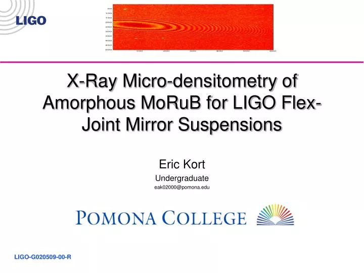
X-Ray Micro-densitometry of Amorphous MoRuB for LIGO Flex-Joint Mirror Suspensions Eric Kort Undergraduate eak02000@pomona.edu LIGO-G020509-00-R
Overview • Gravity waves, LIGO, and Suspensions - quick overview • X-Ray Micro-densitometry - why it is necessary • The Process - how x-ray micro-densitometry is done • Results - what we were and will be able to certify with this technique Laser Interferometer Gravitational-Wave Observatory
Gravity waves, LIGO, and Suspensions Einstein: Gravity is described by warping in space-time. http://www.npr.org/programs/atc/features/2002/sept/gravitywaves/index.html Gravity Wave: Ripple created in fabric of space-time that propogates at the speed of light. (caused by things such as super nova explosions and the big bang) Laser Interferometer Gravitational-Wave Observatory
Gravity waves, LIGO, and Suspensions LIGO will (hopefully) detect these gravity waves, directly confirming GR and opening a new realm of astronomy http://www.ligo-wa.caltech.edu/aerial_full.jpg Detectionis done through a Michelson Morley laser interferometer Gravity waves cause mirror displacement, resulting in length changes in different directions in each arm, producing signals for us to interpret Laser Interferometer Gravitational-Wave Observatory
Gravity waves, LIGO, and Suspensions However Gravity Wave displacement very small (~10-18m!!!) (proton’s diameter ~10-15m) Need to hang the mirrors very carefully Currently- piano wire Predicted upgrade- fused silica wires Better upgrade?- amorphous metal flex joints Laser Interferometer Gravitational-Wave Observatory
Gravity waves, LIGO, and Suspensions Amorphous MoRuB currently being manufactured and tested here at Caltech Looks Promising!!! Current Plan of flex-joint shape 300 microns long, 3mm wide, and 10 microns thick Plan to hang tens of kilogram, fraction of a million dollar mirrors off these joints Laser Interferometer Gravitational-Wave Observatory
X-Ray Micro-densitometry Must be sure joint is correctly constructed How? • Series of tests verifying material properties (stress/strain etc..) • X-Ray Diffraction (determines glassiness) • X-Ray Micro-densitometry Bragg Jr. Laser Interferometer Gravitational-Wave Observatory
X-Ray Micro-densitometry 4 Incentives for X-Ray Micro-densitometry to determine the uniformity of the thickness, compactness, and density of the splat-quenched sample (splat-quenching- technique used to make glassy metals) to certify the absence of cracks or holes (larger than the critical defect size) in the splat-quenched sample to select a region of the splat-quenched sample suitable to flex-joint creation (glassy and flat) to certify the final flex joint is uniform, has the desired shape, and is defect free Laser Interferometer Gravitational-Wave Observatory
Incentives Incentives Splat-Quenched Sample Each splatted sample is unique Need to know information on each sample to pick a region good for testing and, eventually, good for a flex joint (Thanks to Brian Emmerson for the photo) Laser Interferometer Gravitational-Wave Observatory
How does it work?The Process 3 Main Stages: X-Ray imaging the sample (thanks to the animal care facility for their help and the use of their machines) digitizing the image (thanks to the digital media center for their assistance and the use of their machines) analyzing the image in Matlab Laser Interferometer Gravitational-Wave Observatory
The Process:X-Ray Imaging We used a standard diagnostic X-ray unit (just like what the doctor uses on someone when they break a finger, or what a vet would use to see the babies in a pregnant lemur) and standard mammography film by adjusting power settings we could image our sample so we could see thickness fluctuations, cracks etc.. (thanks to Dr. Russel Rose and Dr. Virginio Sannibale for help with this x-ray session) Color enhanced x-ray Laser Interferometer Gravitational-Wave Observatory
Digitizing used Kodak Slide Scanner (4000ppi, translates to pixel dimensions of ~6x6 microns) Analysis done in Matlab production of color maps, thickness-intensity number corrolation, pixel neighbor corrolation test, 2-d profiles The Process:Scanning and Analyzing Laser Interferometer Gravitational-Wave Observatory
Results Cut strips Whole splats OK Can select suitable region to cut Can re-asses cut region Bad Bad Want Uniform Strips Laser Interferometer Gravitational-Wave Observatory
Results Post Electro-chemically polished sample (thanks to Stefano Tirelli for poloshing the sample) 45 5 Crystals are eaten preferentially, leaving holes which are evident is x-rays, indicating impure sample Laser Interferometer Gravitational-Wave Observatory
Quantitatively • Can Detect Defect down to 6x6 microns (critical defect size ~100 microns) • Locally can resolve surface variations ~4 microns (large scale ~27 microns) Laser Interferometer Gravitational-Wave Observatory
Conclusions Success Have technique which fulfills 3 of 4 designated tasks • Determine Uniformity • Locate Cracks/Holes • Identify Amorphous Regions 4th Task, characterize finished flex joint, is expected to be a straight-forward development Laser Interferometer Gravitational-Wave Observatory
Acknowledgments Thanks to: Dr. Riccardo DeSalvo, Hareem Tariq, Animal Care facility, Digital Media Center, Dr. Russel Rose, Professor Johnson, Dr. Jan Schroer, Stoyan Nikolov, Professor Francesco Fidecaro, Virginio Sannibale, Mike Hall, Yoichi Aso, Kelin Wang, Brian Emmerson, Stefano Tirelli, and the rest of Riccardo’s group. Caltech, LIGO, the LIGO REU program, and the SURF office. And, of course, the patient animals that waited in line for their X-rays. Laser Interferometer Gravitational-Wave Observatory