Download
1 / 45
730 likes | 2.18k Views
Fat Embolism Syndrome. Dr. S. Parthasarathy MD., DA., DNB, MD ( Acu ), Dip. Diab . DCA , Dip. Software statistics PhD ( physio ) Mahatma gandhi medical college and research institute, puducherry , India. History .

E N D
Fat Embolism Syndrome Dr. S. Parthasarathy MD., DA., DNB, MD (Acu), Dip. Diab. DCA, Dip. Software statistics PhD (physio) Mahatma gandhi medical college and research institute, puducherry, India
History • In 1861, Zenker described fat droplets in the lung capillaries of a railroad worker who sustained a fatal thoracoabdominal crush injury. • In 1873, Bergmann was first to establish the clinical diagnosis of fat embolism syndrome.
What is it ?? • complex with potentially catastrophic cardiopulmonary and cerebral dysfunction • Three problems : • dyspnoea, petechiae and mental confusion
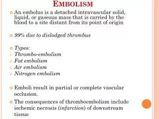
Definitions Fat Emboli: Fat particles or droplets travel through the circulation Fat Embolism: fat emboli passes into the bloodstream and lodges within a blood vessel. Fat Embolism Syndrome (FES): serious manifestation of fat embolism occasionally causes multi system dysfunction, the lungs are always involved and next is brain
Fulminant fat embolism • sudden intravascular liberation of a large amount of fat causing pulmonary vascular obstruction, severe right heart failure, shock and often death within the first 1-12 h of injury
Trauma related (95 %) • Long bone fractures • Pelvic fractures • Fractures of other marrow-containing bones • Orthopaedic procedures • Soft tissue injuries (e.g. chest compression with or without rib fractures) • Burns • Liposuction • Bone marrow harvesting and transplant
Non-trauma related • Pancreatitis • Diabetes mellitus • Osteomyelitis and panniculitis • Bone tumourlysis • Steroid therapy • Sickle cell haemoglobinopathies • Alcoholic (fatty) liver disease • Lipid infusion • LAST OPD – pneumonic
What is frequent ?? • lower extremity and pelvic trauma, • intramedullarynailing of long-bone fractures, • hip arthroplasty, and knee arthroplasty
Incidence ?? • incidence of FES was 1 % • But multiple fractures, adults, high velocity injuries, cementing, hypovolumia • It can be upto 33 %
Lethal dose • The acute lethal dose of fat ranges from 20-50 ml. • The volume of marrow fat from a femur is approximately 70-100 ml. • Mortality – 10 – 20 %
Pathophysiology ?? • The Mechanical theory (Gauss) • Biochemical theory (Lehmann and Moore) • Coagulation theory
The Mechanical theory (Gauss) • Trauma to long bones releases fat droplets • (10-40 μm in diameter) • fat droplets enter the torn veins near long bone ( intramedullary pressure is higher than the venous pressure) • They enter lungs • perivascularhemorhage and edema- picture of ARDS • but smaller ones ( 7- 10 mic.) travel to systemic circulation via ? Patent foramen ovale -
Biochemical theory • Embolized fat is degraded in plasma to free fatty acids. • FFA can cause lung injury, cardiac contractile dysfunction • CRP appears to be responsible for lipid agglutination and may also participate in the mechanism of non-traumatic FES.
Coagulation theory • Tissue thromboplastin is released with marrow elements following long bone fractures. • Activates intravascular coagulation • fibrin and fibrin degradation products, leukocytes, platelets and fat globules combine to increase pulmonary vascular permeability • Catecholamines are involved
Sickling • Bone marrow necrosis as a result of hypoxia may release fat
Number of theories means • Poorly understood ??
Clinical Features • 12-72 hrs after the initial injury • Rarely two weeks
Features • Respiratory changes – 95 % • Cerebral changes – 60 % • petechiae (33% - 60 %). • Not necessary to follow one by one
Respiratory changes • Dyspnoea, tachypnoea and hypoxaemia are the most frequent early findings. • Respiratory failure as ARDS
Cerebral • The more common presentation is with an acute confusional state • but focal neurological signs including hemiplegia, aphasia, apraxia, visual field disturbances have been described. • Seizures and decorticate posturing have also been seen. • Fortunately, almost all neurological deficits are transient and fully reversible.
Petechiae • Embolization of small dermal capillaries leading to extravasation of erythrocytes. This produces a petechial rash in the conjunctiva, oral mucous membrane and skin folds of the upper body especially the neck and axilla • No relation to platelets • Self limiting (36 hours to seven days)
Petechiae Neck
Petechiae • Petechiae only rarely appear on the legs and they are never seen on the face or the posterior aspect of the body. WHY ?? • May be – • fat globules float and therefore distribute to branches of the aorta that arise from the top of the arch, and to the side of the body that is uppermost
Gurd – 1 major + 4 minor • Major – • Axillary or subconjuctivalpetechiae • PaO2 < 60 with FiO2 of > 40 • CNS depression disproportionate to hypoxemia • Pulmonary edema ( PODE – Pneumonic) • Minor • tachycardia, pyrexia, retinal fat emboli, (Purtscher’s retinopathy )urine or sputum fat, Increased ESR, Decreased platelet/ hematocrit. • exclusion of other posttraumatic causes of hypoxemia • Beware a lung injury
How to confirm ?? • High index of suspicion and some investigations
CXR usually normal early on, later may show ‘snowstorm’ pattern- diffuse bilateral infiltrates
Lab values • Arterial blood gases : • This reveals a low partial pressure of oxygen and a low partial pressure of CO2 with respiratory alkalosis. • An unexplained anemia (70% of patients) and thrombocytopenia (platelet count <1,50,000 mm-3 in up to 50% of patients. Hypocalcemia (due to binding of free fatty acids to calcium) and elevated serum lipase have also been Reported Hypofibrinogenemia
CVS • ECG :sinus tachycardia ; Non specific ST T changes, RBBB, • Lung scan : ? V/Q mismatch. • Transesophageal echocardiography : Fat droplets. PFO, Rt sided dilatation if present
Broncho alveolar lavage • BAL : fat droplets. • The staining of cells with oil red O after recovery by a standard 150- to 200-mL lavage can identify intracellular fat droplets. • Can be there in minimal fat embolism – but!! • quantitative count of lavage cells containing fat of greater than 30% being significant of fat embolism syndrome
CT Brain • White matter petechiae • Cerebral edema • Rarely cerebral atrophy due to • full embolisation
Treatment • Prevention and supportive • adequate oxygenation and ventilation, • stable haemodynamics, • blood products as clinically indicated, hydration, • prophylaxis of deep venous thrombosis and stress-related gastrointestinal bleeding, • Nutrition care
Prevention • Hole and drill the long bones • Early immobilization of fractures • Cementless prostheses or • bone-vacuum cementing technique • Less reaming • Albumin also binds fatty acids and may decrease the extent of lung injury • Methylprednisolone 1.5 to 7.5 mg / kg IV 6 to 12 doses (depending on the risk) ?? Advantage
Prevention • during cementing • Hydration • Oxygenation • No nitrous
Treatment • Aspirin • Heparin • N acetyl cysteine • Other speculated therapies such as glucose and insulin, alcohol infusion therapy have theoretical benefit • Details of mechanical ventilation, Inhaled nitric oxide, inhaled prostacyclins – not covered
Prognosis who survived • The prognosis for patients who survive fat embolism is good, with recovery from the fat embolism syndrome usually being complete within 2-4 weeks. • neurological signs may remain for up to 3 months
Summary • Definitions • Incidence • Etiology • lethal dose • Theories • Prevention • Treatment