Download
1 / 73
760 likes | 957 Views
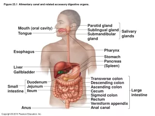
Figure 23.1 Alimentary canal and related accessory digestive organs. Figure 23.2 Gastrointestinal tract activities. Figure 23.3 Peristalsis and segmentation. Figure 23.3a Peristalsis and segmentation. Figure 23.3b Peristalsis and segmentation.

E N D
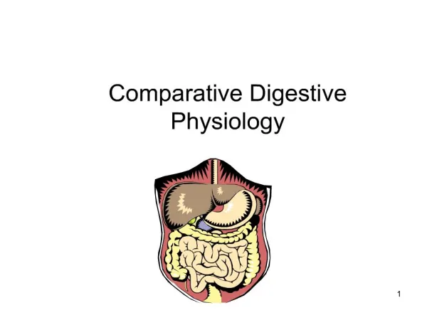
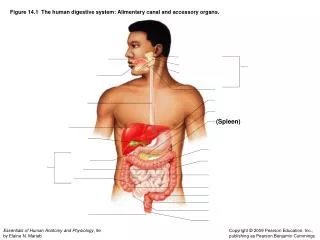
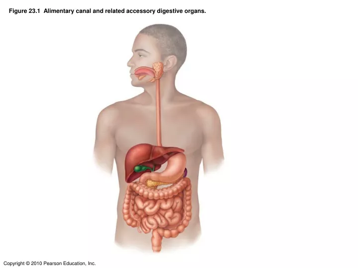
Figure 23.1 Alimentary canal and related accessory digestive organs.
Figure 23.4 Neural reflex pathways initiated by stimuli inside or outside the gastrointestinal tract.
Figure 23.11 Longitudinal section of a canine tooth within its bony alveolus.
Figure 23.16 Photographs of a gastric ulcer lesion and of the bacteria that most commonly cause it.
Figure 23.16a Photographs of a gastric ulcer lesion and of the bacteria that most commonly cause it.
Figure 23.16b Photographs of a gastric ulcer lesion and of the bacteria that most commonly cause it.
Figure 23.17 Neural and hormonal mechanisms that regulate release of gastric juice.
Figure 23.20 Neural and hormonal factors inhibiting gastric emptying.
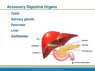
Figure 23.21 The duodenum of the small intestine, and related organs.
Figure 23.22a Structural modifications of the small intenstine that increase its surface area for digestion and absorption.
Figure 23.22b Structural modifications of the small intenstine that increase its surface area for digestion and absorption.
Figure 23.22c Structural modifications of the small intenstine that increase its surface area for digestion and absorption.