Download
1 / 36
360 likes | 866 Views
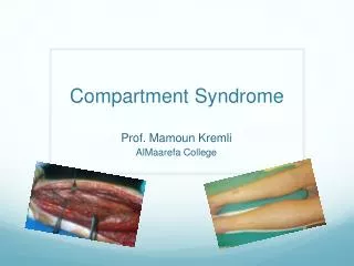
The Painful Truth about Compartment Syndrome, Causalgia, and Post-traumatic Pain Syndromes. Sharla Owens, M.D. Vascular Conference November 14, 2005. Compartment Syndrome. Defined as increased tissue pressure contained in a nonexpansile space

E N D
The Painful Truth about Compartment Syndrome, Causalgia, and Post-traumatic Pain Syndromes Sharla Owens, M.D. Vascular Conference November 14, 2005
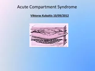
Compartment Syndrome • Defined as increased tissue pressure contained in a nonexpansile space • Raised pressure within a closed fascial space reduces capillary perfusion, placing the enclosed structures at risk • Most commonly observed after acute injury or ischemia in upper and lower extremities • Abdominal, epidural, CHI, and angle-closure glaucoma are better defined and treated as variants of classic compartment syndrome
Pathophysiology • Mechanism of: tissue volume increasing within a confined space, available tissue space decreasing, or a combination of both • Space-occupying lesions such as hematoma, abscess, PSA, muscle edema/fluid • Decreased space by circumstances where external devices are placed (cast, dressing) or with circumferential burns
Ischemia-Reperfusion • Ischemia results in depletion of intracellular energy stores, which, after reperfusion will generate toxic oxygen radicals. • Results in cascade of activation of leukocytes/platelets, inflammatory mediators, calcium influx, disruption of cell membrane, and fluid transudation • All culminate in excess fluid formation
Inflammatory Mediators • Released from ischemic skeletal muscle • Produce local and systemic effects • Local: increase capillary permeability, activate coagulation cascade to produce more tissue damage • Magnitude of systemic response depends on amount of muscle mass involved
Consequences • Venular pressure, normally 4-7 mm Hg, increases progressively because of venous outflow obstruction from increased tissue pressure • Ultimately, when intracompartmental pressure equals capillary pressure, nutrient blood flow is reduced to 0, cellular perfusion ceases, and tissue infarction commences
Vicious Cycle Increasing capillary pressure Increasing compartmental pressure Fluid transudation and cellular swelling
Clinical Presentation • Most frequently occurs with major extremity trauma (crush or closed fractures) or reperfusion after acute arterial insufficiency • Has also been reported with electrical injuries, massive volume resuscitation, and prolonged operative positioning • Symptoms include severe pain, which worsens despite appropriate care for underlying injury, and neurologic signs
Continued… • Early neurologic dysfunction explained by the fact that tissues most sensitive to hypoxia are nonmyelinated type C sensory fibers (fine touch) and result in symptoms such as paresthesias. • If hypoxia continues, then affects myelinated nerves, skeletal muscle, and then skin and bone. • Muscle cell dysfunction fuels the progression of compartment syndrome
Continued… • Physical findings commonly include tense muscle compartments that are tender to palpation, as well as passive flexion and extension • Numbness and weakness in the distribution of nerves passing through compromised compartments
Objective Tests • Objective measurement of compartment pressure using needles/catheters was developed in the 1970s. Today, many still use needle manometry, and wick catheters • Traditionally, absolute measures were used to guide therapy; pressures > 40-45 mm Hg at any point, or sustained at greater than 30 mm Hg for more than 3-4 hours mandated fasciotomy. • Compartment syndrome (CS) occasionally develops at lower tissue pressures in hypotensive patients
Objective Tests, cont’d. • Isolated measurement of compartment pressure is neither sensitive nor specific for determining muscle ischemia • Important variable is the gradient between diastolic blood pressure and compartment pressure
Objective Tests, cont’d. • CS cannot exist without derangements of venous flow dynamics • Jones and coworkers determined that venous duplex scanning focused on tibial veins might be an accurate means of indirectly determining presence of compartmental pressures.
Objective Tests, cont’d. • Subsequent work by Ombrellaro showed that loss of normal respiratory venous phasicity correlates well with elevated tissue pressures. • The finding of normally phasic tibial venous flow on duplex scanning could effectively rule out elevated tissue pressures in that compartment.
Objective Tests, cont’d. • Additional noninvasive modalities include fiberoptic transducers, near-infrared spectroscopy, laser Doppler flowmetry, and tissue tonometry • Above approaches are experimental, and such data should not be used independently to exclude the presence of compartment syndrome
Treatment • Reactive oxygen species play a key role in the development of compartment syndrome by producing DNA damage, which triggers complex energy consuming DNA repair mechanisms, leading to more inflammatory mediators, and potentially cell death. • Pretreatment with mannitol, catalase, superoxide dismutase, or other scavengers, has been shown in models to diminish cell damage after I-R injury
Treatment, cont’d. • Although these experimental observation have implications as adjunctive therapies, surgical decompression, or fasciotomy remains the mainstay of therapy. • Fasciotomy is indicated when compartment pressure is within 20-30 mm Hg of diastolic pressure • Sole purpose is to release compartment HTN and prevent necrosis of compressed tissue
Treatment, cont’d. • In the lower extremity, four compartment fasciotomy of the calf can be performed through a single lateral incision, started one fingerbreadth anterior to fibula and carried out to the lateral malleolus. • Access to anterior, lateral, and superficial posterior compartments is straightforward after raising narrow skin flaps. Access to deep posterior is best obtained distally where the gastrocnemius and soleus muscles become tendinous.
Treatment, cont’d. • Muscle viability is assessed by color, presence of arterial bleeding, and contraction to galvanic (electrocautery) stimulation.(Muscle relaxing anesthetic agents do not interfere with this response). • Muscles that do not contract should be debrided. • Myoglobinuria should be sought and treated with volume expansion, mannitol/loop diuretics as needed, and urine alkalinization by NaHCO3 to prevent renal complications.
Wound Management • Early and complete closure of the fasciotomy wound is a critical step to maintaining a functional limb • Sterile, moist dressings applied until swelling has diminished to allow closure • Closure: delayed primary vs. secondary vs. skin graft vs. flap
Post-traumatic Pain Syndromes The Quick and Dirty
Causalgia • Derived from Greek causos, meaning “heat”, and algos, meaning “pain”. First case reported in early 16th century. • Early reports from American Civil War described incomplete peripheral nerve injury secondary to penetrating trauma with subsequent burning pain, autonomic dysfunction, and “limb atrophy”.
CRPS • Term developed at consensus committee to replace causalgia and RSD. Hallmarks of syndrome include dysfunction and pain of duration or severity out of proportion to what might have been expected from initiating event. • Cause of pain and underlying pathophysiology remain obscure
CRPS • Complex- dynamic/varied presentation, autonomic, cutaneous, motor, inflammatory, and dystrophic changes • Regional- wide distribution beyond the area of lesion; distal part of limb usually affected • Pain- out of proportion to initiating event, hallmark of condition. Refers to spontaneous burning and thermally or mechanically induced allodynia.
Type I (RDS) follows inciting event Pain and allodynia beyond territory of single nerve disproportionate Edema/abnormal skin blood flow in region Type II (Causalgia) results from specific nerve injury Pain and allodynia beyond territory of single nerve Edema/abnormal skin blood flow in region CPRS, cont’d.
Etiology • Traumatic- fractures, dislocations, sprains, burns, crush injuries (Majority of cases) • Nontraumatic- prolonged bed rest, neoplasms, metabolic bone disease, MI, CVA (15% of pts s/p MI develop persistent pain in the UE during their recovery) • Idiopathic
Pathogenesis • Not well understood • Most popular theory is that of “artificial synapses” occurring at site of injury, where a short circuit occurs at the point of partial nerve interruption that allows efferent sympathetic impulses to be relayed back along afferent somatic fibers. • Stimulation of a sensory nerve may make the nerve more sensitive to the usual types of sensory stimuli. • This theory has difficulty explaining similar pain without over nerve injury (CRPS I)
Presentation and Diagnosis • Stage I, acute: warmth, erythema, burning, edema, hyperalgesia, hyperhidrosis, patchy osteoporosis. Good result with chemical sympathectomy, spontaneous resolution possible. • Stage II, dystrophic: Coolness, mottling of skin, cyanosis, brawny edema, brittle nails, diffuse osteoporosis. Good response to sympathetic block, symptoms present for fixed interval, spontaneous resolution rare.
Presentation and Diagnosis • Stage III, atrophic- Pain always extends beyond area of injury, florid trophic changes, atrophy of skin, fixed joint contractures, severe demineralization and ankylosis on plain films. • Diagnosis of CRPS II certain when clinical presentation includes superficial burning pain in distribution of single somatic sensory nerve, hyperesthesia, vasomotor abnormalities, radiographic evidence of osteoporosis, and a good response to local sympathetic blockade.
Diagnostic Sympathetic Block • Validity of clinical diagnosis can be strengthened by a positive response to sympathetic block • Patients should quantify the degree of pain relief experienced (e.g., 100%, 50%) • Degree of pain relief is an excellent predictor of how much relief can be expected from surgical sympathectomy
Treatment • Depends on clinical stage of development, severity of symptoms, and degree and duration of relief by sympathetic block • Disabling pain, several month duration, relief from block for typical duration, should be considered candidates for surgical sympathectomy • Pain of recent onset, with relief from blockade that lasted beyond duration of anesthetic, continue with nonoperative
Treatment, cont’d. • Nonoperative therapy relies on early mobilization for susceptible patients, drug therapy (phenytoin, amytriptyline, and carbamazepine), intermittent sympathetic blocks, and physiotherapy. • TENS (Transcutaneous electrical nerve stimulation) • Steroids • Neuromodulation
Treatment, cont’d. • Stage I: PT +/- TENS, sympathetic block for severe pain or pts unable to undergo PT. If above fails, possible course of steroids. • Stage II: PT, TENS, steroid therapy combined. Sympathetic blocks and surgical sympathectomy considered if measures fail. • Stage III: Steroid or sympathetic block and surgical sympathectomy should be considered, but may be unsuccessful.
Summary • Key to Compartment Syndrome is a high index of suspicion, and early intervention • CRPS- no one really knows what it is, or what causes it, but once diagnosed, the patient should seek therapy, get on medication, and potentially have surgery.