Download
1 / 48
530 likes | 1.33k Views
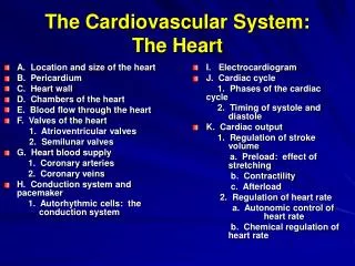
A. Location and size of the heart B. Pericardium C. Heart wall D. Chambers of the heart E. Blood flow through the heart F. Valves of the heart 1. Atrioventricular valves 2. Semilunar valves G. Heart blood supply 1. Coronary arteries

E N D
A. Location and size of the heart B. Pericardium C. Heart wall D. Chambers of the heart E. Blood flow through the heart F. Valves of the heart 1. Atrioventricular valves 2. Semilunar valves G. Heart blood supply 1. Coronary arteries 2. Coronary veins H. Conduction system and pacemaker 1. Autorhythmic cells: the conduction system I. Electrocardiogram J. Cardiac cycle 1. Phases of the cardiac cycle 2. Timing of systole and diastole K. Cardiac output 1. Regulation of stroke volume a. Preload: effect of stretching b. Contractility c. Afterload 2. Regulation of heart rate a. Autonomic control of heart rate b. Chemical regulation of heart rate The Cardiovascular System: The Heart
The heart is the center of cardiovascular system. • 1. double pump a. systemic circulation b. pulmonary circulation • 2. 1,800 gallons of blood per day through about 60,000 miles of blood vessels
Heart Location and Size • 1. middle mediastinum • 2. apex vs base
Pericardium • 1. outer fibrous • 2. inner serous a. parietal layer b. visceral layer (epicardium) __________________ • 3. pericardial cavity • 4. pericardial fluid
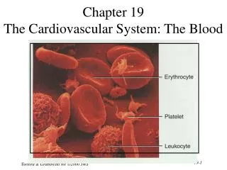
Heart Wall • 1. epicardium • 2. myocardium a. involuntary b. striated c. branched d. intercalated discs e. 2 muscle masses • 3. endocardium (endothelium) Epicardium Fat and connective Tissue + simple squamous epithelium Myocardium Endocardium
Heart Chambers • 1. right and left atrium • 2. atrial appendages • 3. right and left ventricles ____________________ • 4. coronary sulcus • 5. interventricular sulci
Septa of the Heart • 1. interatrial septum a. fossa ovalis b. foramen ovale • 2. interventricular septum a. membranous b. muscular
Heart Valves • 1. atrioventricular (AV) valves (2) a. tricuspid & bicuspid (mitral) b. base vs apex c. chordae tendineae d. papillary muscles • 2. semilunar (SL) valves (2) a. pulmonary valve & aortic valve ______________________ b. 3 half-moon cusps (pockets)
Coronary Circulation(Arteries) • 1. left coronary artery a. anterior interventricular a. b. circumflex a. • 2. right coronary artery a. marginal a. b. posterior interventricular a. _________________________ anastomoses
Coronary Circulation(Veins) • 1. great cardiac v. • 2. middle cardiac v. • 3. small cardiac v. • 4. coronary sinus Small cardiac vein
Conduction System and Pacemaker • 1. heart stimulates itself with autorhythmic cells • 2. ANS and hormones can only modify, not establish, the fundamental rhythm • 3. All cardiac muscle cells are autorhythmic, but 1% lose the ability to contract during early development • 4. those cells that cannot contract form the pacemakers (primary and secondary) and the conduction system
Conduction System Components • 1. sinoatrial (SA) node (90 - 100 action potentials/min) (primary pacemaker) • 2. atrioventricular (AV) node (40 - 50 action potentials/min) (secondary pacemaker) • 3. atrioventricular (AV) bundle • 4. right and left bundle branches • 5. Purkinje fibers
Timing of Conduction Events Times are in seconds 0.01 0.07 0.00 0.09 0.04 0.22 0.05 0.16 0.07 0.19 0.17 0.21 0.19 0.18 0.18 Where does the cardiac muscle contract first? Last? Why is there a 0.12 sec delay across the AV node? What separates the atrial muscle mass from the ventricular muscle mass? Why does the interventricular septum begin to contract before the apex?
Electrocardiogram (ECG) • 1. P wave = atrial depolarization • 2. QRS complex = ventricular depolarization • 3. T wave = ventricular repolarization • 4. P-R interval • 5. S-T segment • 6. quiescent period
Cardiac Cycle Two phenomena control blood flow through the heart. • 1. contraction and relaxation • 2. opening and closing of the AV and SL valves
Blood flows from an area of higher pressure to an area of lower pressure. The pressure developed within a heart chamber is related to two things. • 1. volume of blood within the chamber • 2. size of the chamber
Pressures to Keep in Mind • 1. venous pressure • 2. atrial pressure • 3. ventricular pressure • 4. arterial pressure
In a normal cardiac cycle, the two atria contract while the two ventricles relax, then the two ventricles contract while the atria relax and then all chambers are relaxed until the next P wave • 1. systole vs. diastole • 2. Four phases of the cardiac cycle: a. Ventricular filling b. Isovolumetric contraction b. Ventricular ejection c. Isolvolumetric relaxation
Ventricular Filling • 1. In diastole, ventricles expand and pressure decreases. AV valves open when atrial pr. > ventricular pr. • 2. Three phases of ventricular filling a. rapid ventricular filling b. diastasis c. atrial systole (30%) • 3. end-diastolic volume (EDV) = 130 ml of blood 70% 70% of filling occurs during quiescent period- time period In which all four chambers are in diastole
Isovolumetric Contraction • 1. Atrial diastole begins and Ventricular systole begins • 2. AV valves close when ventricular pr. > atrial pr. • 3. No blood is ejected because arterial pr. > ventricular pr.
Ventricular Ejection • 1. Ventricular pr. > arterial pr. • 2. SL valves open when ventricular pr. > arterial pr. • 3. Rapid ejection, then reduced ejection • Stroke volume (SV) = 70 ml • Ejection fraction = SV/EDV x 100 = 54% • End-systolic volume ESV = 60 ml
Isovolumetric Relaxation • 1. Early ventricular diastole • 2. Ventricles expand, pr. decreases • 3. SL valves close when arterial pr. > ventricular pr. • 4. Isovolumetric because the SL valves are closed and AV valves are still closed The quiescent period begins when atrial pr. > ventricular pr. AV valves open and the period of rapid ventricular filling begins for the next cycle.
Overview of Volume Changes • End-systolic volume 60 ml (left from previous heart beat) • Passively added during atrial diastole + 30 ml (rapid ventricular filling + diastasis) • Atrial systole + 40ml _____________________________________________________________ • total = end-diastolic volume 130 ml • Stroke volume - 70 ml (ejected by ventricular systole) • Leaves the end-systolic volume 60 ml
Timing of the Cardiac Cycle • 1. resting heart rate = 75 beats/minute • 2. one cardiac cycle requires 0.8 sec. • 3. first 0.4 sec. = quiescent period • 4. next 0.4 sec. = atrial and ventricular systole
Chamber Volume and Pressure Changes 8 sec. 0 sec. aortic valve closes aortic valve opens pressure aortic pressure AV valve closes AV valve opens atrial pressure ventricular pressure volume R R ventricular volume P P T T ECG Q Q S S lubb dupp lubb dupp heart sounds ventricular systole ventricular diastole
Cardiac output (CO) = amount of blood ejected from the left ventricle into the aorta per minute • Cardiac output is determined by two factors: 1. stroke volume (SV) 2. heart rate (HR) • So how is cardiac output calculated? CO = SV x HR = (70 ml/beat) x (75 beats/minute) = 5,250 ml/minute or 5.25 liters/minute (L/min)
Cardiac Reserve • 1. difference between maximum cardiac output and resting cardiac output • 2. expressed as percent of normal Normal cardiac reserve = 15-20L/min = 200-300% • endurance athlete = 35L/min = 600%
Regulation of SV: Preload increased venous pressure decreased heart rate increased venous return increased lengthof diastole increased ventricularfilling increasedpreload increased ventricularfilling increased ventricular stretch Frank-Starling mechanism increased force of contraction increased stroke volume increased cardiac output
Regulation of SV: Contractility increased sympathetic activity increased epinephrine other factors increased contractility increased force of contraction increased stroke volume increased cardiac output
Regulation of SV: Afterload increased arterial pressure increased afterload decreased blood volume ejected into artery decreased stroke volume decreased cardiac output
Autonomic Regulation of the Heart • 1. CO = SV x HR • 2. sensory input a. baroreceptors b. chemoreceptors • 3. cardiovascular center • 4. motor output a. cardioacceleratory nerves (sympathetics) b. cardioinhibitory nerves (parasympathetics)
Chemical Regulation of the Heart Rate • Hormones- epinephrine, norepinephrine, throxine, and glucagon increase heart rate • Ions • Increased K+ decreases heart rate • Moderate increase in Ca2+ increases heart rate ,