Download
1 / 78
790 likes | 818 Views
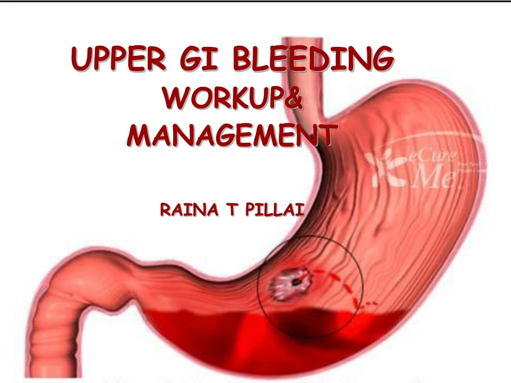
UPPER GI BLEEDING WORKUP& MANAGEMENT RAINA T PILLAI. DEFINITION. BLEEDING EMANATING FROM SITE PROXIMAL TO LIGAMENT OF TRIETZ. HISTORY. SEVERE GI BLEED. Defined as bleed :-- Accompanied by shock or orthostatic hypotension

E N D
UPPER GI BLEEDING WORKUP& MANAGEMENT RAINA T PILLAI
DEFINITION BLEEDING EMANATING FROM SITE PROXIMAL TO LIGAMENT OF TRIETZ
SEVERE GI BLEED • Defined as bleed :-- • Accompanied by shock or orthostatic hypotension • Decrease of PCV by 6 to 8% or transfusion of at least 2 units of packed cells • Bleeding more than 1 litre
Common Causes • Peptic disorders • Duodenal ulcer • Gastric ulcer • Reflux esophagitis • Gastritis • Duodenitis • NSAID associated disorder • Acute gastric mucosal lesions
Other Causes • Portal hypertension-related causes • Esophageal varices • Gastric varices • Portal hypertensive gastropathy • ‘watermelon’ stomach • Mallory weiss tear • Neoplasm of the esophagus, stomach duodenum or ampulla of vater • Swallowed blood
Rarer Causes • Esophagitis due to infection • Dieulafoy’s lesion • Aortoduodenal fistula • Angiodysplasia • Crohn’s disease • Hemobilia • Hemorrhage from a pancreatic source
Clinical manifestations • Hematemesis • Malena • Hematochezia • Occult bleed • asymptomatic
HEMATEMESIS Fresh bright blood or older coffee brown material Signifies larger&brisker upper GI bleeding HEMATOCHEZIA Arise from proximal GI source if 1.Intestinal transit time is fast 2.Bleeding is massive
MALENA - Presence of >50ml blood with transit time >12hrs - Bleeding source proximal to caecum OCCULT BLOOD - slow or intermittent bleed not evident to the patient - may present with features of anaemia Occassionally NO signs of GI bleed – only HYPOVOLEMIA
CLINICAL FEATURES DEPEND ON • Rate • Nature of blood loss • Duration • Amount • Active bleeding • Co-morbid conditions
HISTORY 0F PRESENT ILLNESS • a/c hematemesis, malena or hematochezia • upper abdominal pain • recent dyspepsia/heartburns • recent violent retching/vigorous coughing • Previous upper GI bleed
(Contd…) • Family/personal h/o nosebleed • Aortic surgeries • Pancreatitis • Heavy alcoholconsumption • Aspirin/NSAID intake
EXAMINATION • First examine patient’s vital signs >40% blood vol. loss-shock -systolic B.P.in supine pos.<90mm of Hg -tachycardia -cold extremities 20- 40 %blood vol. loss - hypotension in upright pos.
Around 20% blood vol. loss -orthostatic variation in vital signs *>20 beats/min.&decrease in sys.BP >10mm of Hg -signs of peripheral hypoperfusion *cool clammy pale extremities
FIRST PRIORITY- RAPID ASSESSMENT OF PATIENTS HEMODYNAMIC STATUS NOTE: • Patient’s appearance • Pulse rate • Blood pressure • Peripheral perfusion
1.RAPID RESTORATION OF INTRAVASCULAR VOLUME • Insert two large bore(16G) I/vcanula • Immediate blood sample for • Hematocrit • Coagulation parameters • Typing & crossmatching • LFT& RFT
FLUIDS FOR RESUSCITATION In all patients Patients in shock with GI bleed Ringer lactate sol. Prompt transfusion of packed red cells Ongoing haemorrhage Continuous resuscitation with saline & RBC transfusion
2.ADMINISTRATION OF OXYGEN through - nasal cannulae - mask
3.ENDOTRACHEAL INTUBATION Protection of airways -High risk of aspiration *In massive hematemesis *In mental obtundation *Prior endoscopy/balloon tamponade
4. MONITORING • In Massive haemorrhage • Elderly patients • Significant co-morbidities -cardiac -pulmonary -renal -hepatic insufficiencies
MONITOR Vital signs • Urine output with Foley catheter • Central venous/Pulmonary artery catheters -Cardiac & renal diseases -Brisk/ongoing bleeding -High risk of rebleed -Hemodynamic vitals unreliable (elderly & patients on beta-blockers) • Continous ECG • Pulse oximetry • Mental status
5. ADDITIONAL LAB EVALUATION • Complete blood count • Platelet count • Prothrombin time • Partial thromboplastin time • S.Electrolytes • S.creatinine • LFT • CXR
6.MAINTENANCE OF FLUIDS COAGULATION DEFECTS CORRECTION • Component therapy • Fresh frozen plasma(2U of FFP for 4 U of blood) when PT>20 sec 3. Platelet transfusion (Count<50,000) 4. Cryoprecipitate(when fibrinogen<0.2 g/l)
If Vitals return to normal I/v infusion reduced to maintenance rates If patient shows NO response to rapid infusion of 2L of crystalloids Early transfusion of blood products (esp. if hematocrit 30% or less)
CRITERIA FOR ADMISSION BLEED RISK CLASSIFICATION SCHEME Based on 5 criteria: 1.Ongoing bleeding 2.Sys. B.P<100 mm of Hg 3.Prothrombin time>1.2 times control 4.Altered mental status 5.Unstable co-morbid d/s requiring ICU admission Presence of any one – 3 times higher risk of rebleeding
IN THE WARD • High dependency care • Nil per orally & volume replacement • Continuous Ryles tube aspiration • Oxygen inhalation • Serial monitoring • Hematocrit • Input output chart • Continuous ECG • Pulse oximetry
Initial measures • Lavage using ice cold saline &few drops of adrenaline -Reduces bleeding by vasoconstriction -Reduces chance of encephalopathy by washing out stomach contents & blood • Instillation of Sucralfate via Ryles tube (30 ml) • Stop NSAIDS & other offending drugs • Start Antacid therapy (H2 blockers, PPIs)
INVESTIGATIONS • UPPER GI ENDOSCOPY - Investigation of choice - Fully flexible endoscope
Appearance of ulcer at endoscopy - most important predictor of re-bleeding • FORREST CLASSIFICATION SYSTEM
Diagnostic Angiography • Acute and massive(600ml/hour)bleed • Active hmrge of at least 0.5ml/min at the time of angiography • Chronic and recurrent bleed • Vascular abnormalities • Tumor neovascularity • Angiodysplasia • pseudoaneurysm
Video endoscopy Barium studies Endoscopic USG CT (…contd)
BLEEDING PEPTIC ULCER TREATMENT OPTIONS • Medical management • Endoscopic procedures • Surgery
DRUGS USED IN BLEEDING PEPTIC ULCER • H2BLOCKERS-Cimetidine,Ranitidine etc. • PROTON PUMPINHIBITORS-Omeprazole,Pantoprazole • PROSTAGLANDINS-Mifepristol • SOMATOSTATIN • SOMATOSTATIN ANALOGUES-Octreotide • ANTIFIBRINOLYTICS-Tranexaemic acid
ENDOSCOPIC HEMOSTASISINDICATIONS • Actively but not massively bleeding ulcer • Ulcer with stigmata of recent hemorrhage • Bleeding/non-bleeding ulcer with visible vessel • Presence of visible clot
ENDOSCOPIC HEMOSTASIS-OPTIONS • ENDOSCOPIC INJECTION/SCLEROSIS • ENDOSCOPIC CAUTERY • ENDOSCOPIC HEAT PROBE • ENDOSCOPIC LASER
Indications for surgery • Severe bleed unresponsive to initial resuscitation • Hemodynamic instability • Prolonged bleed with loss of half/more of blood vol. • Continued bleed demanding > 1 unit blood every 6 to 8 hrs • Persistent hemorrhage>48 hrs • Rebleed after initial control