Download
1 / 30
400 likes | 1.26k Views
Biomechanics of the skeletal muscles. Skeletal muscles. Are the prime movers of the human body. The forces they produce under the control of the nervous system act on the bones to which they are attached to create the propulsive forces necessary for human movement.

E N D
Skeletal muscles • Are the prime movers of the human body. • The forces they produce under the control of the nervous system act on the bones to which they are attached to create the propulsive forces necessary for human movement. • Man has 640 skeletal muscles of many shapes and sizes from the tiny stapedius muscle of the middle ear to the massive hip extensor, the gluteus maximus). • Muscles are situated across joints and are attached at two or more points to bony levers. • Each muscle is well adapted to provide an appropriate range, direction and force of contraction to meet the habitual requirements at the articulations over which it passes.
Properties of Skeletal Muscles • Irritability : is the ability of the muscle to respond to stimulus. • Contracility: is the capacity of the muscle to produce tension between it’s ends. • Relaxation: is the opposite of contraction and is the giving up of tension. • Both contraction and relaxation progress from zero to maximal values over a finite time. • Distensibility: is the ability of the muscle to be stretched or lengthened up to a certain limit by an outside force; e.g. pull of an antagonist muscle, of gravity or by an opponent. The muscle suffers no harm so long as it is not stretched beyond its physiological limits. • Elasticity: is the ability of the muscle to recoil to its original length when an outside force is removed unless it has been overstretched.
Factors Influencing Muscle Function Shape and Fascicular Architecture Contraction type Myoglobin contents and fiber type Muscle Function Physiological cross section Muscular attachment Interaction in joint movement Relation to joint structure and muscle length Number of joints traversed
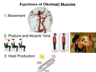
Function of the skeletal Muscles • Create the propulsive force responsible for human movement and positioning of the bony segments of the body. • Give shape to body segments. • Form supportive walls.
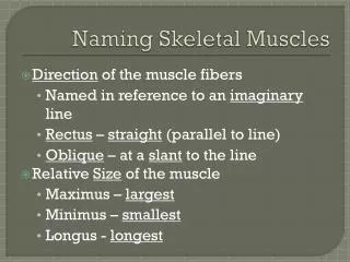
Classification of Muscles: • According to the shape and fascicular architecture: • Parallel: spindle. • Oblique: e.g. pinnate. • Spiral: Supinator. • The muscles designed for strength are of pinnate type and the ones designed for speed have parallel fibers.
2)According to the myoglobin content: • Red: contain more red fibers and they are responsible for movement, which require slow action for a long time e.g antigravity muscles. • White: contain more white fibers and they are responsible for movement, which require rapid action for a short time. 3) According to the type of contractile activity: a. Tonic muscles (stabilizers): it demonstrates continuous low level of contractile activity which is required to maintain a given posture. b. Phasic muscle (mobilizers): it demonstrates rapid (fast twitch) activity which is required when changing from one position to another.
4) According to general limb appearance: • Contractors: those muscles pull the body into approximation of the fetal position e.g. flexor adductors and medial rotators. • Expanders: those muscles which expand or open up the body e.g. extensors. Abductors and lateral rotators. 5) According to the relative magnitude of their stabilizing and rotatory components (muscle attachments): • Spurt: mainly rotator muscles which have their origin away from the joint and their insertion near to the joint e.g. biceps muscle. • Shunt: mainly stabilizer muscles which have origin near the joint and their insertion away from the joint e.g. brachio-radialis.
6) According to the orientation of the line of pull to the joint structure: ( e.g. flexors, extensors, abductors and adductors) • The muscle located anterior to a joint may be extensor as in the case of the knee joint or may be flexor as in the case of the elbow joint. • The possible axes of motion are determined by the structure of the joint itself. 7) According to the number of joints over which the muscle crosses: • One joint muscle ( e.g. vastus mediales). • Two joint muscle( e.g. rectus femoris). • Multi-joint muscle ( e.g. finger flexors).
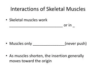
8) According to the type of muscle action or function (their interaction in joint movement): • One action of the joint is not only the responsibility of one muscle but it is the responsibility of different groups of muscles, which can be classified as follows: • Agonists, antagonists, synergists, fixators.
Types of muscle actions or functions: • Agonists: agonists are the muscles which contract to perform a certain action and they include: • Prime movers: Muscles which make the major contribution in any contraction ( e.g. iliopsoas in hip flexion movement). b) Secondary movers: Muscles which cross the same joint but make less contribution in the movement. They are also called accessory or assisted movers. They act sometimes as prime movers when the force required increases or paralysis occurs (e.g. Sartorius in hip flexion).
Antagonists: They are muscles which oppose the prime movers as they relax and lengthen progressively to allow agonists to move. Therefore, the movement is controlled but not impeded. For every action, there are agonists and antagonists (e.g. Gluteus maximus is antagonist for iliopsoas). • Synergists: • Synergists are muscles that work together in a close cooperation as they either contract or relax to modify the action of the agonist. Their aims are: • To make the agonist stronger • To eliminate the action of undesired movement. They may alter the direction of pull and that depends on their power in relation to the agonist muscle.
Types of Synergists: • Conjoint. • Neutralizer. • Stabilizer. a) Conjoint: • They are the two muscles acting together to produce a certain movement which neither of them could produce it alone. They are considered as prime movers of agonists and they are parallel to each other. • E.g. tibialis anterior and peroneous tertious work together to produce dorsiflexion.
b) Neutralizer: They are the muscles that neutralize or cancel the undesired action of other muscles of prime movers or secondary movers. This is more apparent in two- joint muscles which pass across more than one joint and they are capable of performing more than one action which are not needed, so the other muscles or neutralizers must contract to counteract the undesired movement. Neutralizers around the target joint: To oppose the undesired action of the prime movers if it crosses bi- or multi-axial joints (e.g. lateral rotators neutralize undesired motion of adductors of medial rotation during hip adduction).To oppose the undesired action of the secondary movers (e.g. internal rotators neutralize the action of sartorius during hip flexion). Neutralizers for undesired motion on another joint in case of two joint muscles. • For example: Contraction of the finger flexors to grasp an object also tend to flex the wrist. The unwanted wrist flexion is neutralized by wrist extensors.
Stabilizers: Stabilizers are the muscles that surround the proximal joint. They contract and become firm to allow distal joint to move smoothly. Their contraction is generally isometric (e.g. the rotator cuff muscles all contribute their opposing tension to support the humeral head against the glenoid fossa when the arm is moved away from the body and the hand reaches for an object). • Fixators: Fixators are the muscles which contract in both agonists and antagonists simultaneously and that occur especially under stress conditions. • The tension will develop inside both groups of muscles to prevent any degree of freedom. That occurs in normal physiological conditions during strenuous effort and increased demand (e.g. during standing on one leg).
Range of Muscle Extensibility and Contractility: • The maximal degree of angular displacement of a body segment possible at given joints affects all muscles crossing that joint. • The full range of extensibility and contractility of a muscle is called functional excursion or it’s amplitude. • The excursion depends on • the arrangement of muscle fibers and • whether the muscle is a one- joint or a multi- joint muscle with an average magnitude of 57% of their resting length. • Any muscle crossing a single joint is normally capable of shortening sufficiently extensible to permit a full range of motion in the opposite direction.
The absolute amount by which any muscle can shorten depends on: • Length of arrangement of fibers (for pinnate muscles it depends on cos angle of theta of the tendon). • Structure of joint. • Number of joint traversed. • Resistance of antagonists. • Presence of load that oppose the muscle. • Any muscle crossing more than one joint produce motion at the same time in these joints whenever it generates tension up its certain length. • Its efficiency in moving each joint depends on the instantaneous length of the moment arm at each joint and the amount of force that the muscle is exerting for e.g. the rectus femoris muscle is more effective as knee extensor than hip flexor because its moment arm at the hip joint is about 3,4 cm at the knee joint is about 4,4 cm.
The two joint muscles have two different patterns of action which are: • Concurrent pattern occur when simultaneous movement of flexion or extension occur in two joints. • Countercurrent pattern occur when one of the two joint muscles shorten rapidly at both joints its antagonist lengthens correspondingly and thereby gains tension at both ends. • E.g. the rapid loss of tension in the rectus femoris and corresponding gain of tension in the hamstrings when the hip is flexed and the knee is extended simultaneously.
Muscle Insuffisciency: • If a muscle which crosses two or more joints produces simultaneous movement at all of the joints that it crosses, it soon reaches a length of which it can no longer generate a useful amount of tension.
Active insufficiency • I.e. The muscle can not shorten beyond a certain limit without loosing tension and this is called active insufficiency ( e.g. maximal hip flexion with knee extension from a supine lying position).
Passive insufficiency. • When a full range of motion at any joint or joints that the muscle crosses is limited by the anti-agonist muscle length, it is called passive insufficiency. • It is defined as follows: the muscle can not e stretched beyond certain limits without causing pain. (e. g. when a person tries to flex the hip fully with maximal knee extension, he usually feels pain in the hamstring muscle if he has tight hamstrings.
N.B.: When active insufficiency is present in one group of muscles, this does not mean that the opposite group of muscles will suffer from passive insufficiency.
Tendon Action of two joint muscles: • Tendon action is a passive tension without active muscles contraction that may produce movement of the joint if the muscle is elongated over two joints or more than two simultaneously. • E.g. if the wrist is allowed to flex by the weight of the hand the digits will automatically extend without contraction of the finger extensors. By reversing the wrist movement, the fingers will partially flex.
Physiological Cross section of a muscle • The physiological cross section of a muscle determine its potential force of contraction (absolute muscle strength is recognized to be 3-4 kg per sq cm cross section). • Physiological cross section is defined as the area of a section that cut every muscle fibers making up of the muscle and its level of hypertrophy. • It is an indication of the muscle work capacity together with the distance which the muscle can shorten as its functions in the human body : • ( work) = ( force x distance).
Types of bodily movements • Movements can be classified into: • Passive: Subject is relaxed and movement is performed by any outside force. • Active: Is volitionally performed or reflex reaction to an external stimulus. • It is divided into: a. Slow or rapid tension movement that involve constant application of force. b. Ballistic movement: Movement is initiated by vigorous muscular contraction and completed by momentum.
Ballistic movement is terminated by: • Contraction of antagonist muscles. • Reaching the limit of motion and it will be stopped by the passive resistance of ligaments or muscles. • Interference of an obstacle.