Understanding Amoebiasis: Parasitic Infection Overview
320 likes | 559 Views
Explore the taxonomy, morphology, life cycle, clinical aspects, and diagnosis of Entamoeba histolytica, the causative agent of amoebiasis. Learn about its geographical distribution, hosts, and complications.

Understanding Amoebiasis: Parasitic Infection Overview
E N D
Presentation Transcript
AMOEBIASIS By Dr. Gamal Allam Associate Prof. of Immunology & Parasitology
IntroductionScientific Nomenclature • Each parasite has: Phylum , class, order, family, genus & species King Philip Came Over For Good spagetti
Scientific Nomenclature …cont • The scientific name of the parasite is binomial: Genus& species • The Genus starts with a Capital letter and species name starts with small letter • The scientific name of the parasite should written in Italic letter or Under lined.
Human Parasites Parasites infect human fall into 2 categories: 1- Unicellular Parasites (Protozoa). 2- Multicellular Parasites (Metazoa).
Classification of human parasitic protozoa • Kingdom: Protozoa 1- Phylum: Rhizopoda • Entamoeba 2- Phylum: Ciliophora • Balantidium 3- Phylum: Euglenozoa (Mastigophora or Flagellates) • Trypanosoma • Leishmania 4- Phylum: Apicomplexa (Sporozoa) • Toxoplasma • Plasmodium
Learning outcomes By the end of the lecture, you should be able to: Mention systematic position of Entamoeba histolytica. Mention geographical distribution, habitat & hosts of Entamoeba histolytica. Describe morphology of Entamoeba histolytica. Explain life cycle of Entamoeba histolytica. Mention pathology and clinical complication of Amoebiasis Diagnose Amoebiasis. Differentiate between E. histolytica and E. coli.
Suggested Reading • Chiodini, P.L.; Moody, A.H. and Manser, D.W. (2001): Atlas of Medical Helminthology and Protozoology. 4th ed. Churchill Livingstone, P. 48-53. • http://www.dpd.cdc.gov/dpdx/HTML/Para_Health.htm • http://www.dpd.cdc.gov/dpdx/HTML/Amebiasis.htm
Phylum: RhizopodaClass: Entamoebidea e.g.Entamoeba histolytica
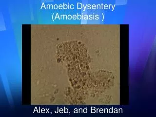
Entamoeba histolyticaDisease:Amoebiaisis, amoebic dysentery, amoebic colitis, amoebic liver abscess. Geographical distribution: Cosmopolitan especially in tropics and subtropics and wherever sanitary conditions are bad.Habitat: Large intestine, occasionally extra-intestinal (liver, lung, brain, …).Hosts: Definitive host: man. Reservoir host: monkey, dog and rat.
MorphologyThree stages [Trophozoite - precyst - cyst]1- Trophozoite: Size: (average 20 µm).It has clear ectoplasm and granular endoplasm with food vacuoles containing RBCs in the invasive forms.The pseudopodium is well developed with active, progressive, motility
2- Precyst:Spherical or oval with single pseudopodium & sluggish movement.Smaller than the trophozoite but larger than the cyst.Has single nucleus.Has no food inclusions in the cytoplasm.3- Cyst:- Spherical, 10 – 20 µm (average 15 µ) with one, two or four nuclei.- The nuclei resemble that of the trophozoite.- Cigar-shaped chromatoid bodies and diffuse glycogen mass are present in young cysts and represent stored food
Life cycle: Infective stage: quadrinucleated mature cyst. Mode of infection: ingestion of cysts:1- In contaminated food and drinks. 2- By autoinfection.3- By hand to mouth through direct faeco- oral contamination from person to person especially among family members.
Ingestion of mature cyst Excystation in the small intestine Metacyst with 4 nuclei multiplication 8 separate metacystic trophozoites from one cyst. The trophozoites proceed downstream to colonize the lumen of the colon and multiply by binary fission. Life cycle …. cont
Important points Definitive host: human Reservoir host: monkey, dog and rat Infective stage: quadrinucleated mature cyst Diagnostic stage: Cyst & trophozoite Mode of infection: Ingestion of mature cyst
Clinical aspect *Asymptomatic infection. The infected persons are usually healthy carriers who excrete millions of cysts / day without any clinical symptoms. Very dangerous as a source of infectionand spread. *symptomatic infection: 1- Intestinal Amoebiasis A- acute dysentery (diarrhea alternating with constipation, tenesmus with blood & mucucs in stool). B- chronic non-dysenteric amoebiasis. 2- extra-intestinal amoebiasis: The trophozoites may disseminate via blood to other extra-intestinal sites e.g. in the liver, lung, brain … etc.
Extraintestinal amboebiasis • *Hepatic amoebiasis: • Amoebic hepatitis: sudden rise of temperature + enlarged tender liver • Amoebic liver abscess: Fever, pain in the right hypochondrium that usually refers to the right shoulder and enlarged tender liver. If not treated the abscess may rupture and trophozoite may go to pleural cavity, lung, peritoneal cavity, pericardium, gall bladder and skin. Aspiration of the abscess yields anchovy sauce (thick chocolate-coloured pus) with trophozoites. *Amoebic lung abscess
Direct Extension Haematogenous spread Extra-intestinal Amoebiasis Perianal amoebiasis
Diagnosis • A- Clinical: clinical picture and endemicity. • B- Laboratory: I- Intestinal amoebiasis: Direct: 1-Stool examination by: -Direct smear: Cyst or trophozoite can be detected in stool. Trophozoite appears more in diarrheic stool while cysts are present more in well-formed stool -Concentration techniques as zinc sulfate-flotation may be needed when cysts are few.
Diagnosis …. cont • 2-Stool culture. • 3-Rectal scraping: to detect trophozoites. • 4-Sigmoidoscopy or total colonoscopy for: Visualization of the lesions- Biopsy- Aspiration.
Diagnosis …. cont • Differences between E. histolytica & coli • E. Histolytica E. coli Pathogenic commensal non pathogenic- • Trophozoite 10-20μm , 20-30μm. • Mature cyst 4- nucleated 8- nucleated
Diagnosis …. cont II- Extra-intestinal amoebiasis: • X-rays. • Ultrasonography. • Computed tomography (CT) and magnetic resonance imaging (MRI). • Immunological tests. • Examination of aspirates for trophozoites by smear or culture. • Leucocytic count: leucocytosis.
Prevention and Control • Environmental sanitation: • Anti-vector measures. • Proper sewage disposal. • Safe water supply. • Not to use excreta as fertilizer or storage before use. • Health education: • Washing of green raw vegetables. • Washing hands before eating and after defecation. • Treatment of carriers, particularly food handlers.