Download
1 / 23
230 likes | 779 Views
ACQUIRED COAGULATION ABNORMALITIES. ACQUIRED COAGULATION ABNORMALITIES - causes. 1. Vitamin K deficiency 2. Liver disease 3. Clotting factor inhibitors : a) circulating anticoagulants b) complications of anticoagulant therapy

E N D
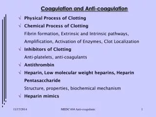
ACQUIRED COAGULATION ABNORMALITIES - causes 1. Vitamin K deficiency 2. Liver disease 3. Clotting factor inhibitors : a) circulating anticoagulants b) complications of anticoagulant therapy 4. Incraesed consumption or loss of the clotting factors: a) disseminated intravascular coagulation ( DIC) b) fibrinogenolysis (primary fibrinolysis)
Coagulation abnormalities vitamin K deficiency dependent • vitamin K is essential for the final postribosomal carboxylation of coagulation factors II, VII, IX, X and the physiologic anticoagulants, protein C and protein S • laboratory features - prothrombin time (PT) is prolonged and decreased factors II, VII, IX, X level • activated partial thromboplastin time (aPTT) - prolonged in severe, protracted vitamin K deficiency
Vitamin K deficiency-etiology I. Inadequate supply: 1. Dietary deficiency 2. Destroying the gut flora by administration of broad-spectrum antibiotics II. Impaired absorption of vitamin K: 1. Biliary obstruction (gallstone, strictures, tumor) 2. Malabsorption of vitamin K(sprue, celiac disease, ulcerative colitis) 3. Drugs (cholestyramine) III. Pharmacologic antagonists of vitamin K (coumarins, warfarin)
Abnormalities of hemostasis and coagulation in liver diseases (1) I. Decreased synthesis of coagulation factors 1. Fibrinogen, protrombin, clotting factors V, VII, IX, X, XI, XII, XIII, prekallikrein, high molecular weight kininogen 2. Antiplasmins, antithrombin, protein C and S II. Aberrant biosynthesis : 1. Of abnormal fibrinogenu 2. Of abnormal analogues of prothrombin, factors VII, IX, X,
Abnormalities of hemostasis and coagulation in liver diseases (2) III. Deficient clearance : 1. Of fibrin monomers, fibrinogen degradation products (FDP) 2. Of activated coagulation factors (IXa, Xa, Xia) 3. Of plasminogen acivators IV. Accelerated destruction of coagulation factors: 1. Intravascular coagulation 2. Localized coagulation (hepatic cell necrosis) 3. Abnormal fibrinolysis V. Thrombocytopenia and platelet dysfunction (splenomegaly)
Circulating anticoagulants Clotting factor inhibitors are autoantibodies (usually IgG) or alloantibodies ( in hemophilia A) that inactivate coagulation factors and, therefore, act as anticoagulants - The laboratory test: prolonged aPTT,
Circulating anticoagulants I. Antibodies to factor VIII (prolonged aPTT, normal INR) 1. In hemophilia A 2. Postpartum -several months after parturition in asociation with a first pregnancy 3. Various immunologic disorders (rheumatoid arthritis, SLE, penicillin allergy ) 4. Older patients without underlying disease II. Other spontaneous inhibitors (rarely)- against factors : V, IX, XIII, fibrinogen, III. Lupus anticoagulant (in 30% SLE, rheumatoid arthritis, HIV infection, in lymphoproliferative disorders, after drugs hydralazine, quinidine, penicillin)
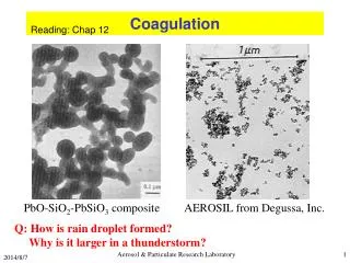
Disseminated intravascular coagulation - DIC DIC is an acquired syndrome characterized by the activation of intravascular coagulationup tointravascularfibrin formation.,The process may be accompanied by secondary fibrinolysis or inhibition of fibrinolysis. This definition implies that microclot formation and consecutive organ failure and/or a hemorrhagic diathesis may occur.
DIC- ETIOLOGY 1. Infections: viral (herpes, acute hepatitis), bacterial (meningococcemia, septicemia), mycotic (aspergillosis), protozoal (malaria), 2. Abruptio placentae, septic abortion, postpartum hemolytic-uremic syndrome 3. Neoplasms: carcinomas (prostate, pancreas, lung, ovary) 4. Disorders of hematopoietic system:acute leukemia (promyelocytic ) intravascular hemolysis 5. Vascular disorders: giant hemangiomas, aneurysmas, myocardial infarctioncardiac arrest, various forms of shock, 6. Massive tissue injury: large traumatic injuries and burns, 7. Miscellaneous: acute pancreatitis, graft versus host disease, diabetic acidosis,
ACUTE DIC-CLINICAL PRESENTATION • symptoms of underlying disease • symptom of local thrombosis • hemorrhagic diathesis • shock
Diffuse intravascular coagulationMicrothrombosis secondary fibrinolysis ↓ platelets FDP clotting factors Ischemic tissue damage Microangiopathic Bleeding anemia tendency
AcuteDIC-laboratory features • Increased D-Dimer level • Increased FDP level • Decreased AT level • Decreased platelet level • Bload smear - schistocytes • Decreased fibrinogen level • Prolonged thrombin time • Prolonged aPTT • Prolonged prothrombin time (PT)
Acute DIC diagnosis The basis of the diagnosis is the knowledge of the underlying diseases in which DIC can occur. Because patients suffering from acute DIC need urgent therapy, DIC should always be taken into consideration if a complex coagulation defect in combination with a underlying disease is observed.
CHRONIC (compensated) DIC In chronic DIC, the activation of the hemostatic system is minimal since negative feedback mechanisms as well as inhibitors can limit the activation process so that microthrombi do not occur and bleeding episodes are rare phenomena.
Chronic DIC - etiology 1. Obstetric complications: eclampsia, the death fetus syndrom 2. Vascular disorders: giant hemangiomas (Kasabach Merrit syndrome), Leriche syndrome, Raynaud,s disease 3. Carcinomas 4. Hematology disorders: myelofibrosis, polycythemia vera, NNH 5. Collagen-vascular disorders: SLE, sclerodermia 6. Kidneys disorders: glomerulonephritis, HUS 7. Another: vasculitis allergica, diabetes mellitus
PRIMARY FIBRINOLYSIS (FIBRINOGENOLYSIS) DEFINITION: primary fibrinolysis occurs when plasmin is generated in the absence of DIC. This has been described in hepatic disorders, prostatic carcinomas, and cases without apparent cause. At present, most cases of primary fibrynolysis are iathrogenically induced during thrombolytic therapy.
FIRINOLYSISPlasminogen intrinsic extrinsic exogenousactivation activation activationfactor XIa, XIIa, kallikrein tPA, uPA streptokinasekininogen or APSAC PlasminFibrinogen Fibrin FDP FDP + D-Dimer
Acquired coagulation abnormalities - diagnostics I History II Physical examination III Laboratory features - morphology - blood smear - bleeding time - prothrombin time (PT), INR - aPTT - thrombin time (TT) - fibrinogen - fibrin(ogen) degradation products (FDP) - D-dimer - antithrombin
Diferentiation of aquired coagulation abnormalities PT aPTT Platelet Fibrinogen TT FDP D-Dimer AT count Acute DIC Chronic DIC N N N N N Fibrinogenolysis N N N N N Heparin overdosage N N N N N NDicumarol N N N N N N overdosage orprothrombin complex factors defficiency
ACA – THERAPY (1) I Vitamin K deficiency - Vit. K i.m. / i.v. 10-20 mg/doba - severe bleeding - Vit.K i.v. + FFP() 10-15 ml/kg II Liver disease - Vit. K - severe bleeding: FFP or prothrombin complex concentrates (FEIBA, Autoplex) III Warfarin overdosage - withhelt anticoagulant on 1-2 d, reduction of dosage - moderate complications – Vit,. K - severe bleeding – FFP or prothrombin complex concentrates, FFP
ACA- Therapy (2) IV Bleeding disorders of circulating inhibitors - factor VIII inhibitor - massive FVIII replacement , cryoprecipitate - FVIII porcin - immunosuppressive therapy - plasmapheresis and high –dose i.v. IgG - activated FVII V Heparin-induced hemorrhage: stop the heparin infusion, intravenous protamin administration at the dosage of 1mg/100U of heparin, FFP
ACA – DIC THERAPY 1. Treatment of the underlying disorder 2. Treatment of shock 3. Replacement therapy - platelet concentrates - RBC - FFP - Cryoprecipitate (fibrinogen) - activated protein C 4. Heparin treatment (unfractioned heparin or low-molecular weight heparin (acrocyanoza, purpura fulminans, dermal necrosis, venous thromboembolism)