Download
1 / 55
580 likes | 1.22k Views
First Trimester Issues. H Murray Dept Obstetrics Nepean Hospital. First Trimester Issues. First trimester bleeding Threatened Miscarriage Ectopic Pregnancy Anti D Genetic screening in the first trimester. Bleeding in early pregnancy (1).

E N D
First Trimester Issues H Murray Dept Obstetrics Nepean Hospital
First Trimester Issues • First trimester bleeding • Threatened Miscarriage • Ectopic Pregnancy • Anti D • Genetic screening in the first trimester
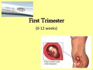
Bleeding in early pregnancy (1) • A woman with no previous pregnancies presents with 6 weeks amenorrhoea, PV spotting and right sided discomfort. Examination is unremarkable other than mild right pelvic tenderness.The HCG is 250IU/L. A pelvic scan shows no intrauterine sac and no other pelvic pathology.
Bleeding in early pregnancy (2) • Which of the following are true? • Rational management includes • A) Referral to hospital as ectopic pregnancy • B) Plan to repeat HCG in 24 hours • C) Plan to repeat US scan in 48 hours • D) Referral to a private specialist requesting urgent laparoscopy • E) Administer anti-D if the woman is Rh negative
Threatened miscarriage/Ectopic • Statistics • 25% of all pregnancies associated with amenorrhoea will fail • 22% of pregnancies with viable fetus at 6.5 weeks will bleed, and 20% of those will fail • A fetal heart at 10 weeks is associated with a miscarriage risk of 3%
Characteristics of viable intrauterine pregnancy • HCG level that doubles every 48 hours • A pregnancy sac that is seen in-utero on ultrasound scan when the HCG is • > 1000 IU/L Transvaginal • > 1800 IU/L Transabdominal • NB Pain is found in 40% viable pregnancies.
Characteristics of viable intrauterine pregnancy • A yolk sac will be seen transvaginally when the gestational sac is > 8mm (5.3 weeks) • A fetal pole should be seen transvaginally when the gestation sac > 20mm (5.5 weeks)
Characteristics of viable intrauterine pregnancy • A fetal heart will be seen with the CRL > 4mm. • The normal fetal heart rate at 6 weeks is 70-100 beats/min.
Implications for scanning • There is no point asking for a scan until the HCG is greater than 1000IU/L • All women must have a full bladder when they present for their scan so that the whole pelvis can be visualised • A transvaginal scan will be necessary to determine fetal viability before 7 weeks • Patients should be warned of this • Patients should be counselled about the safety.
Management of threatened miscarriage • There is no medication that has been proven to be useful in threatened miscarriage in the otherwise normal patient • Specific disorders do have appropriate management • PCOS Metformin/progesterone • Antiphospholipid Aspirin/Heparin • Homocysteinaemia Folate/pyridoxine
Management of threatened miscarriage • Viable pregnancy plus • Bleed settled rescan for NT • Subchorionic haematoma rescan 7-10 days • Continued bleed rescan 7-10 days • Subsequent scan shows POC only consider D&C if endometrial contents > 15mm thickness or heavy bleeding.
Ectopic - characteristics • 2% of pregnancies (50% spontaneously abort) • Presents typically with PV bleed and pain • HCG seldom rises above 6000IU/L • HCG does not double consistently every 48 hours. • May have a pseudodecidual sac in utero in 20% of cases
Ectopic • Ensure good scanning unit • Refer for • High clinical suspicion • HCG > 1000IU/L and no intrauterine sac. • Midcavity pseudodecidual sac • Report of blood in pelvis • Suspicious pelvic mass
Ectopic - management • If mass is < 2cms, tube is unruptured, no active bleeding, HCG < 2000IU/L • Methotrexate 1mg/Kg IM • HCG days 2 & 7. • (Rescan day 5) • Others:- Removal of tube
Anti-D • Which of the following are true about anti-D? • The dose of anti-D in a first trimester miscarriage is 650IU • All Rh neg pregnancies should be given a dose of anti D at 28 weeks • A woman (G2P1) who at an 8 week booking visit has an anti-D titre of 1:1028 should be urgently referred to a hospital clinic • A woman who tested Rh neg at your booking visit is found to be Rh positive at the 28 week visit in the hospital clinic. The difference is likely to be laboratory error.
Background • Sensitisation in Rh –ve women • 1) Transplacental • 75% of women Kleihauer positive in pregnancy • 1st trimester 3%, 0.03 ml • 3rd trimester 45%, 2.5ml • (0.25ml required for sensitisation)
Sensitisation • 2) Blood transfusion • 3) Delivery • 4) Abortion • spontaneous < 1% usually 1ml • therapeutic 20-25% Kleihauer positive
Prevention • No prophylaxis • 16% sensitised • With ABO incompatibility 2% • Prophylaxis • At delivery only:- 1.7% will sensitise • 28 weeks and delivery:- 0.8% will sensitise • 28 and 34 weeks and delivery:- 0.5% sensitise • Manual removal and LUSCS have larger amounts of fetal blood transfer
Common Rh +ve Phenotypes • CcDe • CDE • cDEe • cDe • CcDEe • cDe
Other Rh blood groups • Rh blood groups also have 40 other a/gens • Commonest = Du variant • Cde/cDe Rh+ but the C causes diminished D expression • Tests as Rh negative but is Rh positive • Doesn’t need prophylaxis • Du-positive :- part of the D antigen is missing • Tests as Rh +ve, but acts as Rh -ve • Needs prophylaxis (Anti D)
Anti D - indications • Threatened miscarriage • Miscarriage • Termination of pregnancy • Ectopic pregnancy • Post CVS/Amniocentesis/Amnio reduction • Antepartum Haemorrhage • Trauma with positive Kleihauer • External cephalic versions • Post delivery (amount determined by Kleihauer)
Anti D – New indication • Rhesus negative mother in first term pregnancy • Dose given at 28 and 34 weeks • Needs to have considerable coordination of testing.
Anti D doses • 1st Trimester :- 250 IU Anti D either stat or 3 weekly if bleeding recurrs • 2nd/3rd trimester :- 625 IU Anti D • Used in the 28/34 week dosing • Used for all situations where amount of fetal blood lost is unknown • Used every 3 weeks if bleeding continues.
Anti D doses • 2nd and 3rd trimester • 625 IU dose will cover 4 mls of fetal blood. If the Kleihauer shows more blood lost the dose of Anti D will have to be increased.
Anti D doses • Post delivery • All cases should have had fetal blood group determination and a maternal Kleihauer!! • For the Rh negative mothers with Rh positive babies then the dose of Anti D is • 600 IU Win Rho SDF for every 4 mls of fetal blood in the maternal circulation.
Anti D doses • Summary • 1st trimester 250 IU Anti D • 2nd/3rd trimester 625 IU Anti D • Post delivery 600 IU Win Rho SDF • More than 1 dose will be required for large bleeds, ongoing bleeding.
Anti D issues • The half-life if Anti D is 21 days. Following the administration of Anti D, the laboratory will detect the antibody in routine testing. • It is therefore important to tell the laboratory about the doses of Anti D given when requesting Blood Group status.
Rh negative, pregnant with antibodies • Check paternal Rh status • Refer all patients with Rh positive husbands before the end of the first trimester.
1st trimester Questions • Indicate which of the following are true about nuchal translucency testing • A) The NT scan is performed between 11.0 and 14.0 weeks • B) NT testing cannot be used in a twin pregnancy • C) NT/PAPP-A testing should only be offered to those would would terminate an abnormal fetus. • D) A ‘High – risk’ NT/PAPP-A result indicates a fetal risk of trisomy 21 of greater than 1/200.
The 11-14 week scan • The primary reason for the scan is to assess the nuchal translucency. • Screening test for aneuploidy, cardiac, diaphragmatic and skeletal anomaly • Offered to all women as no risk involved in testing • Many women who do not wish to terminate still wish to be tested • Many women use the test to determine whether to have CVS or lower risk amniocentesis.l
The NT scan • Nuchal translucency • Performed between CRL 45-85mm (11+2-14 weeks) • Must be done at an accredited unit as the risk relates to size, gestation, maternal age. • A poorly performed NT will markedly alter the risk • Accredited units are audited • Accredited units carry the software to calculate risk
The NT scan • Risk of aneuploidy relates to • Size of NT • Age of mother • Gestation of fetus • NT normally less than 2.5mm
NT scan Size of NT, along with maternal age and fetal age will generate • Risk of T21 in fetus • Risk of T18/13 in fetus • And, if NT > 2.5mm:- indicate an increased risk of • XO, triploidy • Fetal cardiac anomaly, skeletal dysplasia, diaphragmatic anomaly • Fetal infection
Down syndrome vs NT • A high risk is taken as > 1/300 • Without additional blood testing the NT, a calculated risk of 1/300 will identify 5% of the population as high risk, in which 70-75% Down syndrome are found • but 150 amnios are required to detect 1 case aneuploidy
Serum screening • To improve the sensitivity for trisomy 21 the NT scan is accompanied by blood analysis for PAPP-A (pregnancy associated plasma protein A) and free HCG • in Down syndrome :- • PAPP-A levels are lower • free HCG levels are raised • Levels are corrected for maternal weight, smoking, and diabetes.
The NT scan • If combined result gives risk of T21 of > 1/300 • Amnio/CVS is offered. • 30 - 39 amnios required for 1 case T21 • Detects 90-92% T21 • If NT result alone gives risk T13/18 > 1/300 • Amnio/CVS offered • If NT > 2.5 mms CVS offered immediately • If chromosomes are normal fetus is scanned for other anomalies
Rate of Invasive Testing • Rate of invasive testing at Nepean has been decimated.
NT screening:- Summary • NT plus PAPP A/free HCG calculate risk of T21 • NT alone determines risk of T13, T18, • NT above 2.5mm is an indicator of possible XO, triploidy and fetal anomaly (heart/diaphragm/long bone anomaly) • Abnormal NT with normal chromosomes needs 15 18 and 26 week scans for fetal anomaly • NT testing alone can be done in nonidentical twins (no blood testing available). No test available for identical twins.
1st Trimester problems • Which of the following about Chorionic villous sampling (CVS) are true • A) CVS is a better option for genetic diagnosis in the 40 yr old than NT/PAPP-A as the NT test will almost always be positive. • B) A CVS result reliably diagnoses fetal aneuploidy in over 99.9% of cases • C) CVS can only be used to diagnose fetal karyotype and genetic/DNA anomalies • D) The advantage of the CVS is that it can be safely used before 10 weeks and therefore ensure a result by 12 weeks gestation.
CVS/Amniocentesis • Diagnostic tests that carry fetal risk • Offered to those at high-risk of fetal aneuploidy/genetic anomaly either following NT testing or due to other factors – • History aneuploidy, extreme maternal age etc.
CVS • Performed after 10.5 weeks (CRL 36mm) • Is a placental biopsy • Procedure is transcervical or transabdominal depending on the placental site • Earlier CVS can result in partial limb loss • Chorion will give information about • Karyotype • Genetics • Rubella/syphylis infection in the pregnancy
CVS for Karyotype • Gives information about the karyotype of the placenta • 1-2% loss rate after the procedure (1% is procedure related • 2-6% mosaicism rate so amniocentesis may be required • Only advantage is the earlier result • May be used with FISH – rapid diagnosis using probes for chromosomes 13,18,21,X,Y. • FISH results may be unreliable therefore
Amniocentesis • Sampling of amniotic fluid • Gives information on • Karyotype • FP • Presence of toxoplasmosis/CMV infection • Detection of fetal Rh status • Detection of Bilirubin in Rh disease • Some enzymes associated with fetal disease
Amniocentesis • Performed after 14 weeks • Earlier has increased risk talipes • No problem with mosacism • Can be used with FISH, but limitations of this technology must be understood. • Quoted loss rate 0.5 – 1% • Benefits:-lower loss rate, no false positive mosacism rate.