Download
1 / 64
800 likes | 1.44k Views
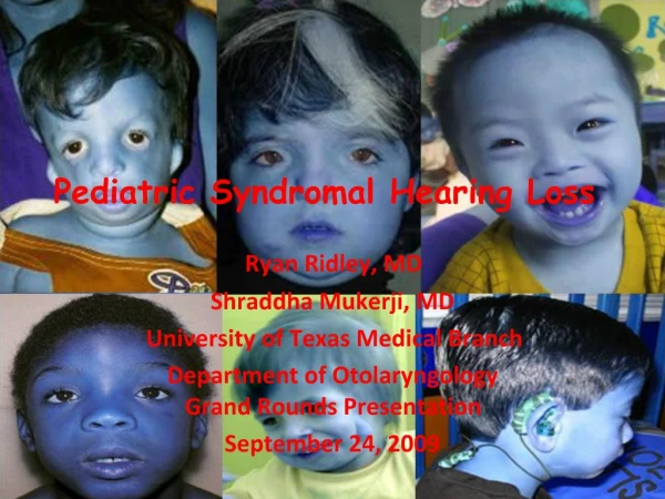
Pediatric Hearing Loss and Testing Techniques. Diego A Preciado MD PhD Pediatric Otolaryngology Children’s National Medical Center George Washington University Washington, DC.

E N D
Pediatric Hearing Lossand Testing Techniques Diego A Preciado MD PhD Pediatric Otolaryngology Children’s National Medical Center George Washington University Washington, DC
11 month child seems to ‘hear well’ at home, but daycare provider concerned with ability to respond to verbal stimuli at times
A) Child can only be tested asleep B) Child can be tested awake C) Child can only be screened for hearing loss at his age D) Child can only be tested by the old ‘rub/snap your fingers’ next to the ear trick
Childhood Hearing Loss Moderate to profound congenital hearing impairment occurs in 4 per 1000 live births Recommendations specify that All children screened at birth All children diagnosed by 3 months of age All children treated by 6 months 43 states have mandated Universal Newborn Hearing Screening (UNHS) programs
Types of Hearing Tests • Screening (PASS OR FAIL) (electrophysiological) • Otoacoustic Emissions (OAE) • Automated ABR • Diagnostic (NOT PASS OR FAIL) • ABR (electrophysiological; all ages) • Pure Tone Audiometry (>4 yrs of age) • Infant Audiometry (>6 mo of age)
Here’s the clinical challenge we are faced with….. Language behaviors UNHS Infants with HL ENT HA, Rehab, SLP Optimal Age for CI Target Age age in months 0 6 12 18
Electrophysiologic Testing • Use in neonates, uncooperative patients, brain injury • Otoacoustic emissions (OAEs) • Originates in cochlea, evoked with sound stimulation • Absent suggests > 30 dB HL
Electrophysiologic Testing • Evoked auditory brainstem response (ABR or BAER) • Auditory electrical responses • Diagnoses presence, degree and type of HL
Hearing Loss Types • Conductive • measured by “air” stimulation on audiogram • Sensorineural • measured by “bone” stimulation on audiogram • Mixed
Conductive Loss • Conductive loss (CHL) results from increase in impedance (resistance) • Audiometric profile of conductive hearing loss is threshold for air conduction is worse than for bone conduction i.e. large “air-bone gap”
Conductive Hearing Loss • Generally reversible • Middle or External Ear Pathology • External auditory canal (EAC) obstruction • Cerumen impactions, foreign body, otitis externa, EAC atresia • Abnormality of ear drum • Perforation, retraction • ME conditions • AOM, OM with effusion, cholesteatoma, tumor • Ossicular chain anomalies • Disruption - associated with trauma • Fixation – often congenital
Tympanometry • Not a hearing test! • Objective measure of middle ear (ME) compliance • Complements ear exam
Tympanograms Volume COMPLIANCE/Admitance
Tympanograms Type A- Normal
Tympanograms Type B- High Volume Perforation or PE tube Type B- Low Volume Fluid
Tympanograms Type C- Negative Pressure Retraction
Sensorineural Loss • Sensorineural hearing loss (SNHL) results because of lesions in the auditory nerves and/or cochlea • Audiometric profile of sensorineural hearing loss demonstrates air conduction and bone conduction reduced without an air-bone gap
70% recessive 25% dominant 5% X-linked Etiology of SNHL
SNHL – Associated Conditions • Loop diuretics, aminoglycosides, aspirin • Hyperbilirubinemia • Severe depression at birth (asphyxia) • Anomalies of external and middle ear • Usually irreversible • Family history – congenital, delayed onset childhood SNHL • Congential infections – CMV, rubella • Bacterial meningitis
Mixed Hearing Loss • Mixed hearing loss results from BOTH a conductive and sensorineural hearing loss • Audiometric profile shows a drop in air and bone conduction with an air-bone gap
Behavioral Audiometry Test Techniques: • Behavioral Observation Audiometry (BOA) • Visual reinforcement audiometry (VRA) • Conditioned play audiometry (CPA) • Conventional hand-raising procedures
Behavioral Observation Audiometry (BOA) • Children aged ~5 months to 2 years • Individuals with neurological/developmental involvements • Primarily sound field testing • Subjective observation by the clinician • Stimuli may include speech, warble tones, narrowband noise (NBN), parent’s voice
Audiometry during infancy Symbols Sound field S S S S Speech = 20 NBN or warbled tones = 20-50 S Age-appropriate responses for infants aged ~5 – 9 months
Visual reinforcement audiometry (VRA) Employs lighted transparent-boxed toys to reinforce child’s localized response to onset of acoustic stimuli
Visual reinforcement audiometry (VRA) • Children aged 6 mo - ~ 3 years • Technique consists of conditioning & testing phases • Responses may include localizations or BOA responses • Disadvantages • Dependent on conditioning child to task • Habituation to acoustic stimuli • Poor test reliability
Conditioned Play Audiometry (CPA) Child is taught a play task in response to the onset of an acoustic stimulus Children aged ~ 2-2.5 - 5 years
Audiometry • Conventional audiometry: ≥ 5 yrs • Zero-20 dB is normal range • Not absolute, but normalized scale • Hearing threshold measured for air and bone conduction in decibels from 250 Hz – 8 KHz
Pure Tone Audiograms ‘loudness’
AudiogramsBracket = Bone, Right Side Circle = Air, Right Side NORMAL
AudiogramsBracket = Bone, Right Side Circle = Air, Right Side CHL A-B gap
AudiogramsBracket = Bone, Right Side Circle = Air, Right Side SNHL
AudiogramsBracket = Bone, Right Side Circle = Air, Right Side MIXED HL
A 3 year old child presents with low volume, Type B tympanogram, and 20 dB Air Bone gap Most likely diagnosis is A- Cholesteatoma B- TM perforation C- OM with effusion D- Sensorineural hearing loss
A 3 year old child presents with low volume, Type B tympanogram, and 20 dB Air Bone gap Most likely diagnosis is A- Cholesteatoma B- TM perforation C- OM with effusion D- Sensorineural hearing loss
Hereditary Hearing Impairment • Dominant • progressive, milder, late onset, penetrance/expressivity • Recessive • stable, severe, congenital, more symmetric
Waardenburg Syndrome • Autosomal dominant • Variable expressivity • Associated with pigmentary abnormalities • White forelock (20-30%) • Premature graying • Vitiligo • Heterochromia irdis
Treacher Collins Syndrome (Mandibulofacial Dystostosis) • Inheritance: Autosomal dominant with variable expressivity • Molecular basis:Caused by mutations in Treacle gene (TCOF1)
Branchiootorenal Syndrome • Autosomal dominant • Branchial cleft sinuses/fistulas • Renal anomalies • These range from mild hypoplasia to bilateral renal agenesis • External, middle and inner ear deformities • Estimated at about 2% of childhood deafness
Pendred Syndrome • Autosomal recessive • Abnormal incorporation of iodine • Perchlorate or thiocyanate tests are rarely performed • Goiter and hypothyroidism usually present by about 8 years of age
Pendred Syndrome • Associated with enlarged vestibular aqueduct • Histologic evidence of hydrops and degenerated changes of the stria vascularis have been described • Treatment – • amplification • exogenous thyroid hormone
Usher’s Syndrome • Usher’s Syndrome Type I (7 loci-MYO7A) • Autosomal recessive • Severe to profound hearing loss • Absence of vestibular response • Slow progression • Slowly progressive visual field deficits beginning as early as age 9-10
Jervell and Lange-Nielson • Autosomal recessive • Severe-to-profound Bilateral • Cardiac conduction defects • Enlarged T-waves • Prolongation of the Q-T interval • Syncopal episodes • Sudden death