Download
1 / 56
580 likes | 649 Views
Learn about the different types of mediastinal masses, their boundaries, divisions, and common contents. Understand the symptoms, diagnosis, and treatment options for thymomas in this informative guide.

E N D
MEDIASTINAL MASSES Prof MoaathAlsmady
Benign or malignant mediastinal masses can develop from structures that are normally located in the mediastinum or that passthrough the mediastinum during development, as well as from metastases of malignancies that arise elsewhere in the body.
The mediastinum is the region in the chest between the pleural cavities that contain the heart and other thoracic viscera except the lungs • Boundaries • Lateral - parietal pleura • Anterior - sternum • Posterior - vertebral column and paravertebral gutters • Superior - thoracic inlet • Inferior - diaphragm
Divisions of the Mediastinum SUPERIOR MEDIASTINUM Superior - thoracic inlet Inferior - transverse thoracic plane Anterior - sternal angle Posterior - IV disc T4 & T5 INFERIOR MEDIASTINUM Superior – transverse thoracic plane Inferior - diaphragm
Anterior mediastinum • Everything lying forward of and superior to the heart shadow • Boundaries • Sternum, first rib, imaginary curved line following the anterior heart border and brachiocephalic vessels from the diaphragm to the thoracic inlet • Contents • Thymus gland, substernal extension of the thyroid and parathyroid gland and lymphatic tissues
Middle mediastinum • Dorsal to the anterior mediastinum, extends from the lower edge of the sternum along the diaphragm and then cephalad along the posterior heart border and posterior wall of the trachea • Contents • Heart, pericardium, aortic arch and its major branches, innominate veins and superior vena cava, pulmonary arteries and hila, trachea, group of lymph nodes, phrenic and upper vagus nerve
Posterior Mediastinum • Occupies the space between the back of the heart and trachea and the front of the posterior ribs, and paravertebral gutter • It extends from the diaphragm cephalad to the first rib • Contents • Esophagus, descending aorta, azygos and hemiazygos vein, paravertebral lymph nodes, thoracic duct, lower portion of the vagus nerve and the symphathetic chain
Regionalization > Teratoma, Thymus Ectopic Thyroid Adenopathy Adenopathy Bronchogenic Cysts Esophageal Duplication Cysts Neurogenic Tumors Esophageal Duplication Cysts Anterior Mediastinum Middle Mediastinum Posterior Mediastinum > >
Anterior Mediastinum • Lymphoma . • Teratoma • Germ cell tumors • Cystic hygromas • Thymic lesions
Anterior MediastinumLymphoma • Usually older child • Hodgkin’s – 14 yrs • Non-Hodgkin’s – 9 yrs • Often have other symptoms and other adenopathy • Frequently have airway compromise
Symptoms • ●Mediastinal mass effects – Direct involvement or compression of normal mediastinal structures cause a wide range of symptoms. These can include cough, stridor, hemoptysis, shortness of breath, pain, dysphagia, hoarseness, facial and/or upper extremity swelling due to vascular compression (eg, superior vena cava syndrome), hypotension due to tamponade or cardiac compression, and Horner syndrome due to sympathetic chain involvement.
Symptoms • Systemic effects – Systemic symptoms such as fever, night sweats, and weight loss can be present in the case of lymphoma or may be due to a variety of paraneoplastic syndromes, such as myasthenia gravis with thymoma.
THYMOMAS • Thymic lesions account for approximately one-half of all anterior mediastinal masses and can include a range of benign and malignant histologies • Thymomas occur in patients of all ages, with a peak incidence between ages 40 to 60 years. The gender distribution is approximately equal
Thymomas are associated with a variety of paraneoplastic syndromes .The most common is myasthenia gravis, which occurs in approximately 30 percent of patients with thymoma. Furthermore, some patients who are diagnosed with myasthenia gravis will be found to have a thymic mass on imaging. Patients with a thymic mass who have not been evaluated for myasthenia gravis should be tested for anti-acetylcholine receptor antibodies. Computed tomography (CT) of the chest.
THYMOMAS • All thymomas originate from epithelial thymic cells • 4% of them consist of a pure population of epithelial cells • Most have mixed populations of lymphoid cells to a varying extent
THYMOMAS • ~50% - asymptomatic, discovered incidentally on CXR or at autopsy • ~30% local symptoms related with pressure or local invasion: SVC sdr., cough, chest pain, dysphonia, dysphagia • ~20%- 70% associated with an autoimmiune disease: • Myasthenia gravis • Pure red cell aplasia • Polymyosistis • hypogammaglobulnemia
Masaoka Classification-1981 STAGE I Encapsulated tumor with no gross or microscopic invasion TREATMENT Complete surgical excision STAGE II Macroscopic invasion into the mediastinalfat or pleura or microscopic invasion into the capsule TREATMENT Complete surgical excision and postoperative radiotherapy to decrease the incidence of local recurrence STAGE III Macroscopic invasion of the pericardium, great vessels, or lung TREATMENT Complete surgical excision and postoperative radiotherapy to decrease the incidence of local recurrence STAGE IVA Pleural or pericardial metastatic spread TREATMENT Surgical debulking, radiotherapy, and chemotherapy STAGE IVB Lymphogenous or hematogenous metastases TREATMENT Surgical debulking, radiotherapy, and chemotherapy
PROGNOSIS • Benign tumors are noninvasive and encapsulated. • Conversely, malignant tumors are defined by local invasion into the thymic capsule or surrounding tissue. • The Masaoka staging system of thymomas is the most commonly accepted system. • Preponderance of evidence indicates that all thymomas, except completely encapsulated stage 1 tumors, benefit from adjuvant radiation therapy • The prognosis of a person with a thymoma is based on the tumor's gross characteristics at operation, not the histological appearance.
DIAGNOSIS • Chest scan is the imaging procedure of choice . • Thymic enlargement should be determined because most enlarged thymus glands on CT scan represent a thymoma. • CT scan with intravenous contrast dye is preferred • to show the relationship between the thymoma and surrounding vascular structures, • to define the degree of its vascularity, and • to guide the surgeon in removal of a large tumor, possibly involving other mediastinal structures
DIAGNOSIS Biopsy: • If a patient presents with atypical features or is found to have an invasive tumor and is under consideration for induction therapy, obtaining preoperative biopsy is indicated. • The limited anterior mediastinotomy . A thoracoscopic approach for biopsy also can be used
If the tumor is small and appears readily accessible, perform a total thymectomy with contiguous removal of mediastinal fat. • If the tumor is invasive, perform a total thymectomy in addition to en bloc removal of involved pericardium, pleura, lung, phrenic nerve, innominate vein, or superior vena cava. Resect one phrenic nerve; however, if both phrenics are involved, do not resect either nerve, and debulk the area. • Clip areas of close margins or residual disease to assist the radiation oncologist in treatment planning
Chemotherapy • The most common chemotherapy drugs in the treatment of thymoma are: • doxorubicin (Adriamycin, Rubex), • cisplatin (Platinol), • cyclophosphamide (Cytoxan, Neosar), • etoposide (VePesid, Etopophos, Toposar), and • ifosfamide (Ifex, Holoxan). • The common combinations used for the treatment of thymoma include: • cyclophosphamide, doxorubicin, and cisplatin, or etoposide and cisplatin.
Radiotherapy • Adjuvant radiation therapy in completely or incompletely resected stage III or IV thymomas is considered a standard of care. • The use of postoperative radiation therapy in stage II thymomas has been more questionable. • Thymomas are indolent tumors that may take at least 10 years to recur; therefore, short-term follow-up will not depict relapses accurately.
Thymic rebound hyperplasia in an 11-year-old girl with Hodgkin lymphoma.
Germ cell tumor — The mediastinum is the most common location for extragonadal germ cell tumors in adults. Germ cell tumors can be either benign (teratomas, dermoid cysts) or malignant (seminomas, nonseminomatous germ cell tumors). Seminomas are more common than nonseminomatous germ cell tumors. All patients with a mediastinal mass that could be a germ cell tumor should have alpha-fetoprotein (AFP), lactate dehydrogenase (LDH), and beta-human chorionic gonadotropin (beta-hCG) measured prior to any therapy.
Enlarged/ectopic thyroid • — Intrathoracic thyroid tissue typically causes symptoms of shortness of breath or dysphagia The intrathoracic mass is usually continuous with the thyroid gland in the neck; only 2 percent of cases are separate from the cervical thyroid.
Middle mediastinum • Lymphadenopathy — Lymphadenopathy is the most common lesion presenting as a mass in the middle compartment of the mediastinum .The most common causes include lymphoma, sarcoid, and metastatic lung cancer.
Middle Mediastinum • < 2 yrs: Remnants of embryonic foregut (trachea & esophagus) • Pericardial cysts • Lymphadenopathy
Middle Mediastinum Bronchogenic Cyst Can beBronchogenic Bronchogenic Cyst • Can be adjacent to or far away from bronchogenic structures • Usually have respiratory epithelium • Can be large and cause respiratory symptoms Cyst • Can be adjacent to or far away from bronchogenic structures • Usually have respiratory epithelium • Can be large and cause respiratory symptoms • adjacent to or far away from bronchogenic structures • Usually have respiratory epithelium • Can be large and cause respiratory symptoms Esophageal Duplication Cyst • Adjacent to or embedded in wall of esophagus • Can have respiratory or GI epithelium • May either obstruct or erode through esophageal wall • Thoraco-abdominal duplications: orginate near duodenum & jejunum and expand to middle mediastinum (gastric mucosa & vertebral anomalies)
Benign cystic tumor — Cystic masses comprise approximately 20 percent of middle mediastinal masses. • Bronchogenic cysts are the most common cystic lesion and are felt to be secondary to abnormal lung budding during development. Bronchogenic cysts are more common in men. These lesions are sometimes identified because patients have symptoms of substernal pain, cough, recurrent infection symptoms, or dyspnea and are typically located in the right paratracheal region and in the subcarinal location.
Cardiovascular aneurysm or anomaly — A variety of cardiovascular pathologies (eg, thoracic aortic aneurysm, pulmonary artery aneurysm, vascular rings) can present as a mediastinal abnormality.
Esophageal tumor — Advanced esophageal tumors may be identified on imaging as a mediastinal mass; however, the location and symptoms such as dysphagia, weight loss, and occult blood loss are not likely to be confused with lymphadenopathy or other middle mediastinal masses
Imaging Studies • Contrast esophagram
Posterior Mediastinum • Ganglioneuroma • Ganglioneuroblastoma • Neuroblastoma
Posterior mediastinum • Neurogenic tumors — Neurogenic tumors represent more than 60 percent of posterior mediastinal masses. These lesions are classified based upon their neural cell of origin. • ●Schwannomas and neurofibromas are benign lesions that arise from the intercostal nerve sheath and make up 90 percent of adult neurogenic tumors.
●Neuroblastomas and ganglioneuroblastomas are malignant tumors that occur most commonly in children and originate from the sympathetic ganglia. • ●Ganglioneuromas are benign lesions that arise from the sympathetic ganglia and are most common in young adults.
Posterior Mediastinum • Ganglioneuroma • Ganglioneuroblastoma • Neuroblastoma
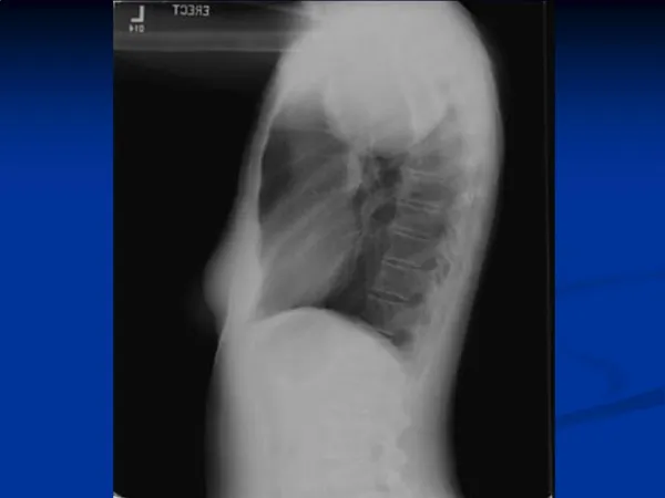
Posterior MediastinumNeuroblastoma • Very good prognosis, especially Stage I & II *Paraplegia implies compression of spinal cord (MRI & urgent laminectomy)
SURGERY • TWO TECHNIQUES: • 1. OPEN MEDIAN STERNOTOMY. • 2. VIDEO ASSISTED THORACOSCOPIC SURGERY ( VATS) • The preferred approach is a median sternotomy providing adequate exposure of the mediastinal structures and allowing complete removal of the thymus,