Download
1 / 15
530 likes | 1.81k Views
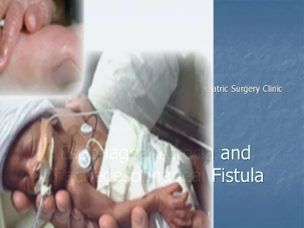
Esophageal atresia. Most frequent congenital anomaly of the esophagus, affecting 1 /4,000 neonates an abnormality in which the middle portion of the esophagus is absent. The esophagus may end blindly into a pouch or may connect to the trachea

E N D
Most frequent congenital anomaly of the esophagus, affecting 1 /4,000 neonates • an abnormality in which the middle portion of the esophagus is absent. The esophagus may end blindly into a pouch or may connect to the trachea • >90% have an associated tracheoesophageal fistula (TEF)
Presentation -Frothing or bubbling of the mouth and nose after birth - Drooling - Choking -Episodes of coughing, cyanosis and respiratory distress
Diagnosis • Prenatal ultrasound- maternal polyhydramnios • inability to pass a nasogastric or orogastric tube in newborn • Plain radiography- shows radiolucent, blind-end dilated pouch of upper esophagus • On lateral view • Anterior displacement of trachea • Rarely, air-fluid level in pouch
Preoperative Management -maintain a patent airway -prevent aspiration of stomach contents or saliva into the lungs -prone position to prevent stomach secretions from refluxing into lungs -Intravenous antibiotics to help prevent the development of pneumonia -The "VACTERL" workup is completed prior to surgical repair
Features • V - Vertebral anomalies • A - Anal atresia • C - Cardiovascular anomalies • T - Tracheoesophageal fistula • E - Esophageal atresia • R - Renal (Kidney) and/or radial anomalies • L - Limb anomalies (in front of or above the central axis of the limb)
Surgical management -Primary anastamosis of proximal and distal esophagus usually when infant is a few months old -gastrostomy, gastric pull-up, colonic transposition and jejunum transposition -
Postoperative care • Postoperatively patients may be extubated as tolerated. • Any attempt at reintubation should be performed carefully in order to avoid accidental and catastrophic intubation of the esophagus. • The head is flexed slightly forward in order to decrease tension on the esophagoesophagostomy. • Oral suctioning performed only to the level of the pharynx. • Endotracheal tube suctioning must be performed carefully in order to avoid trauma or perforation at the site of the TEF closure.
Outcome • Current overall survival rates= 85% - 90% • Mortality is usually secondary to associated anomalies. • Immediate postoperative complications include: • anastomotic leak in 15% of cases. • stricture formation in approximately 15% of cases • recurrent tracheoesophageal fistula in 5% of cases and requires reoperation with division and ligation of the fistula • The most common long term problems : • Gastroesophageal reflux in up to 70% • Tracheomalacia in approximately 25%