Download
1 / 16
200 likes | 502 Views
Implantation. The human oocyte is fertilized in the lateral third of the uterine tube the zygote undergoes cell division as it is moved passively toward the uterus Through successive mitoses, a compact collection of cells, the morula , is formed

E N D
The human oocyte is fertilized in the lateral third of the uterine tube • the zygote undergoes cell division as it is moved passively toward the uterus • Through successive mitoses, a compact collection of cells, the morula, is formed • morula, covered by the zonapellucida, is about the same size as the fertilized oocyte. The cells that result from segmentation of the zygote are called blastomeres • Because the zygote does not grow in size, at each division the blastomeres become smaller.
At the center of the morula a liquid-filled cavity develops and the blastomeres arrange themselves in a peripheral layer (trophoblast) while a few blastomeres accumulate inside the cavity (inner cell mass). • This embryo is now called a blastocyst, which is the stage at which it arrives in the uterus • This happens on approximately the fourth or fifth day after ovulation
In the human, trophoblastic cells over the embryoblast pole begin to penetrate between the epithelial cells of the uterine mucosa about the sixth day • Attachment and invasion of the trophoblast involve integrins, expressed by the trophoblast, and the extracellular matrix molecules lamininand fibronectin. • Integrin receptors for laminin promote attachment, while those for fibronectin stimulate migration. • These molecules also interact along signal transduction pathways to regulate trophoblast differentiation so that implantation is the result of mutual trophoblastic and endometrial action. .
The blastocyst remains in the lumen of the uterus for 2 or 3 days, immersed in the secretion of the endometrial glands, and comes into contact with the surface of the endometrium • The zonapellucida is then dissolved, allowing cells of the trophoblast to interact directly with cells of the uterine surface epithelium. (on the 5th day) • Implantation, or nidation, involves the attachment of the embryo to the endometrial epithelial cells and its penetration into the lamina propria • This type of implantation is called interstitial and occurs in humans and a few other mammals.
The trophoblastiv cells invade the endometrioum by a process of histolysis • It forms a hole by which the blastocyst can enter • The first part of the implant is called embryonic pole • The last part is called the abembryonic pole.
The process starts around the seventh day • At the eighth day of development, the blastocyst is partially embedded in the endometrial stroma. • In the area over the embryoblast, the trophoblast has differentiated into two layers: • (a) an inner layer of mononucleated cells, the cytotrophoblast, • (b) an outer multinucleated zone without distinct cell boundaries, the syncytiotrophoblast
Mitotic figures are found in the cytotrophoblast but not in the syncytiotrophoblast. • Thus, cells in the cytotrophoblast divide and migrate into the syncytiotrophoblast, where they fuse and lose their individual cell membranes. • Cells of the inner cell mass or embryoblast also differentiate into two layers: • a) a layer of small cuboidal cells adjacent to the blastocystcavity, known as the hypoblast layer, • (b) a layer of high columnar cells adjacent to the amniotic cavity, the epiblast layer • Together, the layers form a flat disc.
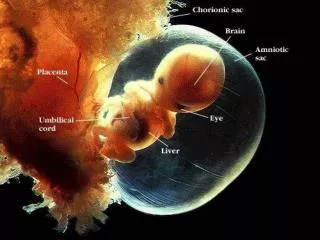
At the same time, a small cavity appears within the epiblast. • This cavity enlarges to become the amniotic cavity. • Epiblast cells adjacent to the cytotrophoblast are calleamnioblasts; together with the rest of the epiblast, they line the amniotic cavity • on about the ninth day after ovulation, the embryo is totally submerged in the endometrium, from which it will receive protection and nourishment during pregnancy. • So the on the 9th or the 10th day the opening is closed by a fibrin clot and the epithelium regenrates • Finally, the whole embryo is embedded in the endometrium. Eventually, the syncytiotrophoblasts come into contact with maternal blood and form chorionic villi. This is the initiation of forming the placenta.
The placenta is a temporary organ and is the site of physiological exchanges between the mother and the fetus • It consists of a fetal part (chorion) and a maternal part (deciduabasalis) • Thus the placenta is composed of cells derived from two genetically distinct individuals. • The deciduabasalis supplies maternal arterial blood to, and receives venous blood from, spaces that exist inside the placenta. • The placenta is also an endocrine organ, producing hormones such as HCG, a placental prolactin, estrogens, and progesterone.
During implantation of the embryo, the endometrial connective tissue goes through profound changes • The fibroblasts of the lamina propria become enlarged and round and exhibit the characteristics of protein-synthesizing cells. • They are now called decidual cells, and the whole endometrium is called the decidua. • Based on the endometrial region, the decidua can be classified as the deciduabasalis, situated between the embryo and the myometrium • the deciduacapsularis, situated between the embryo and the lumen of the uterus • and the deciduaparietalis, the remainder of the decidua • Hence, by the end of the first week of development, the human zygote has passed through the morula and blastocyst stages and has begun implantation in the uterine mucosa
Infertility • Infertility is a problem for 15% to 30% of couples. • Male infertility may be a result of insufficient numbers of sperm and/or poor motility. • Infertility in a woman may be due to a number of causes, including occluded oviducts (most commonly caused by pelvic inflammatory disease), hostile cervical mucus, immunity to spermatozoa, absence of ovulation, and others.
In vitro fertilization (IVF) • In vitro fertilization (IVF) of human ova and embryo transfer is a frequent practice conducted by laboratories throughout the world. • Follicle growth in the ovary is stimulated by administration of gonadotropins. • Fertilized eggs are monitored to the eight-cell stage and then placed in the uterus to develop to term.
Contraceptive Methods • The contraceptive pill is a combination of estrogen and the progesterone analogue progestin, which together inhibit ovulation but permit menstruation. • The intrauterine device (IUD) is placed in the uterine cavity. Its mechanism for preventing pregnancy is not clear but may entail direct effects on sperm and oocytes or inhibition of preimplantation stages of development. • Vasectomy and tubal ligation are effective means of contraception, and both procedures are reversible, although not in every case.
Ectopic pregnancy • In cases of abnormal nidation, the embryo may implant itself in the oviduct (ectopic pregnancy). • In this case, the lamina propria reacts like the endometrium, forming numerous decidual cells • Because of its small diameter, the oviduct cannot contain the growing embryo and bursts, causing extensive hemorrhage that can be fatal if not treated immediately.
Placenta previa • The initial attachment of the embryo usually occurs on the ventral or dorsal walls of the corpus of the uterus • Sometimes the embryo attaches close to the internal os. In this case the placenta will be interposed between the fetus and the vagina, obstructing the passage of the fetus at parturition • This situation, called placenta previa, must be recognized by the physician, and the fetus must be delivered by cesarean section; otherwise, the fetus may die • Sometimes, the embryo attaches to the epithelium of the uterine tube. Very rarely the zygote may enter the abdominal cavity, attach to the peritoneum, and develop there.