
Artemin Modulates Colony Formation and Proliferation in ER-Negative Mammary Carcinoma Cells
This study investigates the role of Artemin (ARTN) in influencing monolayer proliferation and anchorage-independent colony formation in estrogen receptor-negative mammary carcinoma cell lines, BT549 and MDA-MB-231. Monolayer proliferation assays and soft agar colony formation assays were conducted using ARTN-overexpressing and control cells. Cell viability was assessed at multiple time points using Alamar Blue. Results indicate that ARTN promotes increased colony formation in soft agar, highlighting its potential role in the growth of aggressive breast cancer cells.

Artemin Modulates Colony Formation and Proliferation in ER-Negative Mammary Carcinoma Cells
E N D
Presentation Transcript
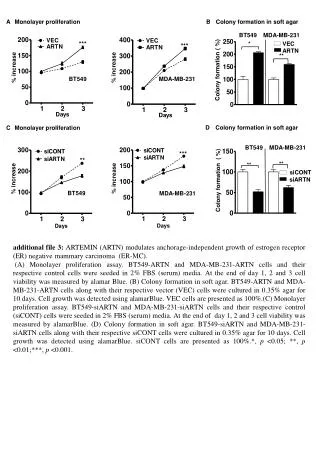
A Monolayer proliferation B Colony formation in soft agar *** ** *** *** BT549 MDA-MB-231 % increase % increase * Colony formation ( %) % increase % increase BT549 MDA-MB-231 ** BT549 MDA-MB-231 D Colony formation in soft agar C Monolayer proliferation siCONT VEC siARTN BT549 MDA-MB-231 ARTN ** ** Colony formation ( %) additional file 3: ARTEMIN (ARTN) modulates anchorage-independent growth of estrogen receptor (ER) negative mammary carcinoma (ER-MC). (A) Monolayer proliferation assay. BT549-ARTN and MDA-MB-231-ARTN cells and their respective control cells were seeded in 2% FBS (serum) media. At the end of day 1, 2 and 3 cell viability was measured by alamar Blue. (B) Colony formation in soft agar. BT549-ARTN and MDA-MB-231-ARTN cells along with their respective vector (VEC) cells were cultured in 0.35% agar for 10 days. Cell growth was detected using alamarBlue. VEC cells are presented as 100%.(C) Monolayer proliferation assay. BT549-siARTN and MDA-MB-231-siARTN cells and their respective control (siCONT) cells were seeded in 2% FBS (serum) media. At the end of day 1, 2 and 3 cell viability was measured by alamarBlue. (D) Colony formation in soft agar. BT549-siARTN and MDA-MB-231-siARTN cells along with their respective siCONT cells were cultured in 0.35% agar for 10 days. Cell growth was detected using alamarBlue. siCONT cells are presented as 100%.*, p <0.05; **, p <0.01;***, p <0.001.