Download
1 / 32
460 likes | 1.15k Views
DIAGNOSIS OF PREGNANCY. ASSOCIATE PROFESSOR IOLANDA BLIDARU MD, PhD. The importance of the problem. avoiding exposure to teratogens (e.g. drugs) solving abnormal situations (e.g. ectopic pregnancy, hydatidiform mole) initial assessment and monitoring through prenatal care. history

E N D
DIAGNOSIS OF PREGNANCY ASSOCIATE PROFESSOR IOLANDA BLIDARU MD, PhD
The importance of the problem • avoiding exposure to teratogens (e.g. drugs) • solving abnormal situations (e.g. ectopic pregnancy, hydatidiform mole) • initial assessment and monitoring through prenatal care • history • clinical examination • laboratory tests Diagnosis The duration of pregnancy in humans • 259 - 294 days =37-42 weeks • medium= 280 days (40 weeks) prezumptive diagnosis During the first half – maternal signs During the second half – fetal signs diagnosisof certitude Diagnosis of pregnancy
Diagnosis ofpregnancy in the first 16 weeks (I-sttrimester) • personaldata • family history • personalmedical history • obstetricalhistory • data about the partener • contraceptive history • generalinformations increased E2 and P secretion by luteal corpus • amenorheea History • Conditions • previous regular, spontaneous, predictible menstruations • absence of menstruation for min. 10 days
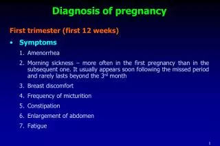
Diagnosis ofpregnancy in the first 16 weeks (I-sttrimester) • enlargement • heaviness or tension • mastodinia, tenderness • Breasts symptoms History • Neuro-vegetative symptoms • nausea ( over 50%) and vomiting • sialoreea • pyrozis • alteration of apetite, taste, smell, food preferences Digestive • appeared during 4-6 weeks • disappeared spontaneously after 12-14 weeks Urinary bladder irritability (compresion) Nervous sleep difficulties, irritability, insomnia
Diagnosis ofpregnancy in the first 16 weeks (I-sttrimester) Clinical signs Inspection • Breasts - increase in size - venous network (Haller) - Montgomery tubercules (sebaceus glands) - minute hyper-pigmentation of the areola and the nipple • Abdomen - linea nigra - progressive deformation upward symfizis pubis (after 12 weeks) • Perineum– accentuation of pigment
Diagnosis ofpregnancy in the first 16 weeks (I-sttrimester) Clinical signs Palpation • Breasts - specific consistency - colostrum (occasionally) – thick, yellowish fluid, expressed from the nipples • Abdomen - pregnant uterus – the upper margin of a globulous, soft mass - the height of the uterine fundus becomes measurable (cm)
Diagnosis ofpregnancy in the first 16 weeks (I-sttrimester) Clinical signs Obstetrical examination Cervix, vagina and perineum (speculum examination) - increased vascularity and hyperemia - characteristic violet-bluish color of the mucosa – Chadwick sign - increased size of the cervix Uterus (bimanual examination) - softening of the cervix, isthmus (Hegar sign) and uterine corpus - enlargement of the uterus (4cm per month) - irregular, painless uterine contractions – Braxton-Hicks contractions
Diagnosis ofpregnancy in the first 16 weeks (I-sttrimester) Noble sign Hegar sign Piscaceck sign
Diagnosis ofpregnancy in the first 16 weeks (I-sttrimester) Differential diagnosis - abdominal mass: ovarian cyst, uterine myoma, trophoblastic disease, ectopic pregnancy, bladder globus - amenorrhea: in the emotional stress, endocrine disfunctions, lactation, anorexia, certain treatments (e.g. antidepressants, metyldopa, haloperidol), pseudocyesis
Diagnosis ofpregnancy in the first 16 weeks (I-sttrimester) Laboratory diagnosis Principle: presence of hCG in plasma and urine - produced by syncytiotrophoblast - peak at 60-70 days - biological tests (laboratory animals) - immunological tests – β-hCG (monoclonal anti-HCG antibody serum assays); home test kit (ELISA, RIA, etc) - detection and quantification
Diagnosis ofpregnancy in the first 16 weeks (I-sttrimester) Ultrasound imaging - abdominal sonography - vaginal sonography can detect an uterine pregnancy 1 week after the missed menstruation AIMS - assessment of the amnionic sac dimensions (4-5 weeks) - visualize the fetus and the placenta (position, aspect) - measure fetal crown-rump length (FCR) - detects twins, ectopic pregnancy, missed abortion, hydatidiform mole.
Diagnosis ofpregnancy in the first 4-5 weeks (I-sttrimester)
Diagnosis ofpregnancy between the 17- 28 weeks (II-ndtrimester) Clinical signs - amenorrhea (> 16 weeks) - progressive enlargement of the abdomen - perception of the fetal movements by the mother beginning with 17-18 weeks (multiparas) up to 19-20 weeks (primiparas) = quickening Inspection • Face: chloasma (melasmagravidarum) = mask of pregnancy • Breasts: increased vascularity, Montgomery tubercules, pigmented primary areola and the nipple, secondary areola • Abdomen: enlargement, linea nigra, stria (reddish, slightly depressed streaks), protruded umbilicus, pigmented scars • Perineum– accentuation of pigment
Diagnosis ofpregnancy between the 17- 28 weeks (II-ndtrimester)
Diagnosis ofpregnancy between the 17- 28 weeks (II-ndtrimester) Clinical signs Palpation • Breasts:specific consistency, colostrum expressed from the nipples • Abdomen: - pregnant uterus – a globulous, soft, contractile, painless mass - irregular, painless uterine contractions – Braxton-Hicks contractions - the height of the uterine fundus becomes measurable (16cm – 20 weeks, 20cm – 24 weeks) - ballottement sign Ascultation • fetal heart sounds (110-160 b/min), with obstetrical stethoscope, near umbilicus
Diagnosis ofpregnancy between the 17- 28 weeks (II-ndtrimester)
Diagnosis ofpregnancy between the 17- 28 weeks (II-ndtrimester) Clinical signs Obstetrical examination – cont. Speculum examination (cervix, vagina and perineum) - hyperemia - Chadwick sign (violet-bluish color of the mucosa) - increased size of the cervix Bimanual examination (uterus) - softening of the vagina, cervix, lower segment and upper segment - enlargement of the uterus - Braxton-Hicks contractions - vaginal ballottement
Diagnosis ofpregnancy between the 17- 28 weeks (II-ndtrimester) Differential diagnosis abdominal mass • ovarian cyst with abdominal development • uterine myoma
Diagnosis ofpregnancy between the 17- 28 weeks (II-ndtrimester) Laboratory diagnosis - biological tests – no more - immunological tests – β-hCG (detection and quantification) Ultrasound imaging abdominal sonography / vaginal sonography AIMS - gestational age (BPD, FL, AC) - fetal morphology and biometry - multiple pregnancy - fetal heart movements - placental insertion, a.f.
Diagnosis ofpregnancy between the 29 - 40 weeks (III-rdtrimester) Clinical signs - amenorrhea (> 29 weeks) - progressive enlargement of the abdomen - perception of the fetal movements by the mother/examinator Inspection • Face: chloasma (melasmagravidarum) = mask of pregnancy • Breasts: increased vascularity, Montgomery tubercules, pigmented primary areola and the nipple, secondary areola • Abdomen: enlargement, linea nigra, striaegravidarum, protruded umbilicus, pigmented previous surgical scars • Perineum– accentuation of pigment
Diagnosis ofpregnancy between the 29 - 40 weeks (III-rdtrimester) Clinical signs Palpation • Breasts:specific consistency, colostrum expressed from the nipples • Abdomen (Leopold manoevers): Superficial palpation - pregnant uterus(a globulous, soft, contractile, painless mass, Braxton-Hicks contractions - measurement of the height of the uterine fundus
Diagnosis ofpregnancy between the 29 - 40 weeks (III-rdtrimester)
Diagnosis ofpregnancy between the 29 - 40 weeks (III-rdtrimester) Clinical signs • Abdomen: Deep palpation - identification of the presenting part (if the head, it is firm, rounded, large, regulated) - lateral palpation – on the sides of the uterus (the back is an elongated firm mass; the limbs are small, irregular parts) Ascultation • fetal heart sounds (110-160 b/min) in vertex presentation – below the umbilicus
Diagnosis ofpregnancy between the 29 - 40 weeks (III-rdtrimester)
Diagnosis ofpregnancy between the 29 - 40 weeks (III-rdtrimester)
Diagnosis ofpregnancy between the 29 - 40 weeks (III-rdtrimester) Clinical signs Obstetrical examination Speculum examination (cervix, vagina and perineum) - hyperemia - Chadwick sign (violet-bluish color of the mucosa) - increased size of the cervix - external cervical os - slit-like or round, with mucous plug Bimanual examination - softening of the vagina, cervix, lower segment and upper segment (patulous cervix admits a fingertip) - fetal presenting part, membranes
Diagnosis ofpregnancy between the 29 - 40 weeks (III-rdtrimester) Ultrasound imaging abdominal sonography / vaginal sonography AIMS - fetal morphology and biometry (BPD, Fl, AC) - fetal heart movements, - breathing movements - evaluation of the amniotic fluid - placental insertion and maturation degree
Presumptive evidence of pregnancy • Subjective symptoms - nausea +/- vomiting - disturbances in urination - fatigue - the perceptions of fetal movements • Presumptive signs - cessation of menses - changes in the breast - changes in the cervical mucus - discoloration of the vaginal mucosa - increased skin pigmentation and development of abdominal striae - does the woman believe that she is pregnant?
Probable evidence of pregnancy • Enlargement of the abdomen • Changes in the shape, size and consistency of the uterus • Anatomical changes in the cervix • Braxton-Hicks contractions • Ballottement • Physical outlining of the fetus • Presence of β-hCG in serum or urine
Positive signs of pregnancy • Identification of fetal heart activity separately and distinctly (from mother) • Perception of fetal movements by the examiner • Recognition of the embryo / the fetus throughout imagistic methods (ultrasound)
Gestational age assessment • First day of the last menstrual period (LMP) + no. of weeks • The day when the mother has felt the first fetal movements (quickening) + 22 weeks in multiparas, or 20 weeks in primiparas • The uterine height (UH) in cm + 4 =the number of gestational weeks • Ultrasound examination during the first 12 weeks of amenorrhea