Download
1 / 51
800 likes | 1.47k Views
Diagnosis of Plant Disease . DR. IR. HADIWIYONO, M.SI. Study Program of Agrotechnology Faculty of Agriculture University of Sebelas Maret (UNS) Surakarta E-mail : hadi_hpt@yahoo.com. Terminologi . Identification:

E N D
Diagnosis of Plant Disease DR. IR. HADIWIYONO, M.SI. Study Program of Agrotechnology Faculty of Agriculture University of SebelasMaret (UNS) Surakarta E-mail : hadi_hpt@yahoo.com.
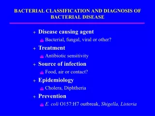
Terminologi Identification: • “the study of the characters of a thing or an organism to determine its name” (APS, 1998) Disease identification: • Study on disease character to determine the name of disease
Diagnosis • Diagnosis is identification of the nature or cause of a disease or other condition • Diagnostic is a distinguishing characteristic important for identification of disease or other conditions (Glossary Plant Pathological Terms, 1998)
Why do diagnosis of disease • For control disease • To plan the disease management in the field • Certification of seed • Plant quarantine to prevent disease spread inter area or countries • For research
Principles of plant disease diagnosis • Simplicity-easy • Times spent-short • Reliability-accurate • Cost need-cheap • Applicability-practical
Standard operational prosedurein identificationand diagnosis of plant diseases • The first step is the determination whether the disease is caused by pathogen or environmental factor • For some cases, the disease symptom and sign appear very obviously or very specific, thus supported by the experience and some available references prove the disease diagnosis to be easy to do. • Most of cases howeverneed detail observation and test to the symptom of diseases.
Diseases caused by parasitic higher plant • The existence of the parasitic higher plant on the host is enough to recognize the causal agent • Benalu (Henslowiafrutescens Champ.) on java apple • Taliputri (Cassythafiliformis L. or Cuscutaaustralis R. Br.) on an ornamental plant.
Diseases caused by fungi and bakteria • While miselia of fungiand spore or ooze of bacterium exhibit in/on the part of diseased plant thus two possibility should be considered: • Fungi or bacterium is the causal disease • Fungi or bacterium is one of many saprophytic fungi or bacterium living on dead tissues caused by other causal agent that is also belong to the group of fungi or bacterium.
Fungi • Some disease have been reported that visual and microscopic observation to structure of mycelia, fruit bodies, and spores associating the symptom followed by checking using relevant manual books or references is enough to determine whether the fungi is the pathogen or saprophyte • In most of cases however, the signs disappear in/on the diseased plant thus identification of the fungi is impossible to do. • Certain fungi must be isolated using selective medium and promoting the sporulation in specific grow chamber.
Method for promoting formation specific structure of fungi in/on diseased tissues • Incubation in high humidity • Incubation under near ultraviolet (NUV)
Method for promoting exhibition of specific structure of certain fungi on pure culture • Incubation under near ultraviolet (NUV) Commonly, the radiation is able to promote the fungi for sporulation 2. Specific treatment i.e. line cutting the medium, enriching with specific substrate, incubating in specific temperature condition.
Isolation and characterization of fungi on culture medium • Structure of colony • Specific structure fungi i.e. forming sclerotium and chlamidospore • Coloring medium by extracellular compound released by the fungi
Chemical and physiological characterization of fungi • The method is less developed • Physiological character of fungi is limited in grow response on the maximum and minimal range of tolerance to salinity, temperature, and pH of medium
Symptom and structure to recognize F.o. f.sp.melonison melon A B C • Discoloration in vascular systems of stem -seem browning • Wilting melons • Microconidia (a), makroconidia (b), clamidospore (c).
Symptom and sign on scallion Alterneriaporrii A. Purple blotch on leaf of scallion; B. Conidia under stereo microscope; C. Conidia under compound microscope
Specific symptom of wilt disease of banana (F.o. fsp.cubense)
Bakteri • Bacterium is too tiny to observe visually or using compound microscope • Specific symptom and sign of disease are often reliable to identification of certain disease in a short time to common diseases being well known • In most of cases, selective medium and specific diagnosis technique are needed
Specific symptom and sign of banana wilt caused by blood disease bacterium (BDB)
Sign of bacterial disease B A A. Sticky strand test on cut stem with bacterial streaming (oozing) from xylem vessels of melon infected Erviniatracheiphila, causing bacterial wilt B. Oozing, bacteria come out from leaf vascular system
Performing bacterium on selective media A. Identification of fluorescence bacteria on King’s B medium; B. BDB on CPG, and C. BDB on TTC medium B A C
Observation of phenotipic colony • Observation the colony structure on specific medium is often used to recognize bacterial pathogen Colony of Xanthomonascampestrispv. campestris, the cause of black rot of cabbage with Yellow colony and forming halo
Hypersensitive Reaction Test • Principle: all of bacterial pathogen exhibit hypersensitive reaction on leaf of tobacco Bacterial suspension 108 sel /mLare injected into the leaf of tobacco (Nicotianatabacum)
Microscopic observation • In severe diseased tissue, the abundance of bacteria can reach 108 – 109 cell/gram of diseased tissue • Washed ooze from diseased tissue or pure culture can observed at 400-1000x • motile bacteria will more observed
Pathogenesity test of bacteria • Pathogenisity test on susceptible host can be employed to diagnose bacterial diseases. • Various inoculation techniques may be applied i.e. • Injection • Spraying on abaxial foliages • - Soil drenching • - Dropping on infection site • - Seed germination on infested medium in plate
Biochemical and physiological test • Well developed and various • Gram reaction test, chemotropic test, capability of carbon degradation, substrate oxidation, nitrate reduction and so forth. • Spend along time and impractical diagnosis but it is a conventional standard characterization in complete identification
Reaction to bacteriophage • Susceptible bacteria to bacteriophagemay be one of specific character of certain pathogenic bacteria Structure of bacteriophage
Diagnosis bacterial disease Commercial automated techniques: Biolog • Spesies bahkan strain memiliki keragaman kemampuan dalam memanfaatkan berbagai sumber karbon • 569 taksa bakteri gram negatif dan 223 gram positif dapat diidentifikasi dengan perangkat ini • Cawan mikrotiter terdiri dari 95 subtrat sumber karbon dan satu kontrol • Inkubasi pada suhu 27-28 oC, selama 4-24 jam • Reaksi terlihat adanya prubahan pewarnaan oleh TTC • Hasil dianalisis dengan perangkat lunak Microlog
Diagnosis bakteri dengan Commercial automated techniques: FAME • FAME (Fatty Acid Methyl Ester Analysis) • Prinsip teknik 1.Bakteri dikulturkan pada kondisi standar 2. Melepaskan asam lemak dari permukaan sel bakteri melalui saponisasi 3. Methylasi asam lemak untuk meningkatkan volatilitas 4. Analisis dengan gas kromatografi resolusi tinggi 5. Membandingkan profil asam lemak yang diperoleh dengan profil mikrobia standar
Identification method based serology (immunology) • High sensitive, specific, accurate, fast, and simple to do • Antiserum monoclonal and polyclonal have been avalable for some plant pathogenic bacteria • Sensitivity 104-106sel/mL
Diagnosis through DNA finger printing • Many method of identification based molecular techniques (Polymerase Chain Reaction) have been developed • Specific primer derived from many plant pathogenic bacteria heve been available • Sensitivities 102-103sel/mL
1 2 3 C- M DNA finger printing based on PCR 1 C+ C- M M 1 2 3 4 • Identification of BDB using primer specific 121F and 121R : 1. BDB, C+. Control-positive, C-. Control negative • Identification Erviniaspp using PCR-RAPD with Universal Primer : 1. E. stewartei, 2. E. carotovora, 3. P. syringe, C-. Kontrolnegatif (Oliver, 1993 • IdentifikasiX.campestrisdengan rep-PCR; 1-2 Xcpvcarotae, 3-4 Xcpvcoriandri A B C
Virus • Virus is obligate parasite, true biotrophic-unculturable • Too small-visualized in light microscope except x bodies belong to some viruses • Identification/diagnosis: specific symptom, pathogenisity test, observation of specific structure in infected cell, specify host test, molecular techniques
Diagnosis of plant diseases caused by virus • For some cases, the symptom of viral diseases are specific. • So using relevant manual book diagnosis is easy to do. • For some other cases, virus diseases cause specific symptom • For other cases, pathogen cause the symptom being similar to other virus infection, tiny pests, toxicity, deficiency, herbicide toxicity and so forth.
pathogenisity test of virus • Some virus can be inoculated mechanically. • Nicotiana spp. and Chenopodium spp. are often used as plant indicator • Type of symptom: local lesion and systemic
A B C Viral diseases with specific symptom A. The symptom of cucumber mozaic viruson lettuce, B. Beet yellow stunt virus on crisp head lettuce (Davis et al. 2002), C. Zucchini yellow mozaic virusonSummersquash (Zitteret al.,1998)
Ex. symptom of pest are similar caused by pathogenic diseases A. Leaf symptom spider mites, B. Spider mites, Tetranychus sp.
Ex. of symptom of herbicide toxicity being similar to virus infection. A. Leaf of squash caused by 2,4-D, B. Glyphosate
Transmitting test • Some plant viruses are insect born • Aphid, Myzus persicae (dewasa) • Whitefly, Bemissia sp. (dewasa) • Aphid, Aphis gossypii (dewasa) • Squash bug, Anasa tristis (nimfa) A B C D
Observation of specific structure of virus in infected cell • Potyviridae produce ‘pinwheel’ inclusions
Diagnosis virus using molecular technique: serological techniques • Virus is enveloped by protein called coat protein • Viral protein (antigen) will act to specific antibody (antiserum) produced by mammalian injected with the antigen • Enzyme-Linked Immunosorbent Assay (ELISA) is the most popular technique • Many antiserum are available in market
Important information addition for disease diagnosis in the fields • Farmer’s name and address • Plant variety • Stage of plant • Pattern of the disease incidence • Insect or other organism • Land history or the previous plantings • Plant environmental condition • Around planting condition • Cultivation practices
Diagnosis of diseases haven’t been well known yet • Certain diseases are well known so the diagnosis are easy to do through observing the symptom and pathogen by then comparing to relevant available manual books or references. • In contrary, the others pathogens may be poorly understood and haven’t been identified yet. For the cases, Koch’s Postulate application is needed.
Koch’s Postulate • Disease agent must be associated with the symptom • The agent must be able to isolate in pure culture • the pure culture inoculated on the susceptible healthy host must be able to induce the symptom on host where the disease agent is isolated • The agent must be able to re-isolate from the inoculated host plant
Standard equipment for diagnosis of plant diseases • Lens or hand Loop • Digital camera with enough resolution • Stereo microscope and compound microscope • Grow or incubation chamber installed lamp and NUV- grow chamber • Isolation room, fungi and bacteria must be separated • Manual book for identification: ex. Compendium, identification guides manuals • Computer with internet on line • KIT for isolation, culture, and microscopic isolation
Conclusions • Disease diagnosis is basic activity being important in plant disease control • Diagnosis of pant disease can be done through: 1. Observation of the symptom and sign 2. Isolation and inoculation on susceptible plant indicator 3. Observation the biochemical and physiological characters of the pathogen 4. Microscopic observation 5. Serological techniques 6. Molecular techniques based on nucleic acid .