Download
1 / 27
280 likes | 478 Views
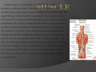
Muscle Soreness. Muscular Soreness. Acute soreness occurs during or immediately following the exercise and is usually caused by ischemia and the accumulation of metabolic waste products in the muscle tissue. Muscular Soreness.

E N D
Muscular Soreness • Acute soreness occurs during or immediately following the exercise and is usually caused by ischemia and the accumulation of metabolic waste products in the muscle tissue.
Muscular Soreness • The pain and discomfort may persist up to 1 hour after the cessation of the exercise.
Muscular Soreness • In delayed-onset muscle soreness (DOMS), the pain occurs 24 to 48 hours after exercise.
Muscular Soreness • Eccentric exercise produces a greater degree of delayed muscular soreness than either concentric or isometric exercise.
Muscular Soreness • The more widely recognized theories suggest that exercise causes damage to skeletal muscle cells and connective tissues, producing an acute inflammation.
Armstrong’s Model (1984) • The structural proteins in muscle cells and connective tissue are disrupted by high mechanical forces produced during exercise, especially eccentric exercise.
Armstrong’s Model (1984) • Structural damage to the sarcolemma alters the permeability of the cell membrane, allowing a net influx of calcium from the interstitial space.
Armstrong’s Model (1984) • Abnormally high levels of calcium inhibit cellular respiration, thereby lessening the cell’s ability to produce ATP for active removal of calcium from the cell.
Armstrong’s Model (1984) • High calcium levels within the cell activiate a calcium-dependent proteolytic enzyme that degrades Z-discs, troponin, and tropomyosin.
Armstrong’s Model (1984) • This progressive destruction of the sarcolemma (postexercise) allows intracellular components to diffuse into the interstitial space and plasma.
Armstrong’s Model (1984) • These substances attract monocytes and activate mast cells and histocytes in the injured area.
Armstrong’s Model (1984) • Histamine, kinins, and potassium accumulate in the interstitial space due to the active phagocytosis and cellular necrosis.
Armstrong’s Model (1984) • These substances, as well as increased tissue edema and temperature, may stimulate pain receptors resulting in the sensation of DOMS.
Acute Inflammation Theory (1991) • Smith suggested that acute inflammation, in response to muscle cell and connective damage caused by eccentric exercise, is the primary mechanism underlying DOMS.
Acute Inflammation Theory (1991) • Connective tissue and muscle tissue disruption occurs during eccentric exercise, especially when the individual is not accustomed to eccentric exercise.
Acute Inflammation Theory (1991) • Within a few hours, neutrophils in the blood are elevated and migrate to the site of injury for several hours post-injury.
Acute Inflammation Theory (1991) • Monocytes also migrate to the injured tissues at 6 to 12 hours post-injury.
Acute Inflammation Theory (1991) • Macrophages synthesize prostaglandins (Series E).
Acute Inflammation Theory (1991) • The prostaglandins sensitize Type III and IV pain affectors, resulting in the sensation of pain in response to intramuscular pressure caused by movement or palpation.
Acute Inflammation Theory (1991) • The combination of increased pressure and hypersensitization produces the sensation of DOMS.
Figure 1. Morphological changes of myofibers after eccentric exercises. A, The control group. B, The 24-hour group. Swollen myofibers (arrowheads) and disrupted myofibers (arrows) are detected. C, The 3-day group. Many myofibers are swollen or disrupted. Small cells (arrowheads) are detected among myofibers. Those cells are probably inflammatory cells and proliferating satellite cells. D, The 7-day group. Myofibers with central nuclei (arrowheads) are detected, which indicates the regeneration of muscle tissue. Hematoxylin and eosin staining. Original magnification is ×200.
Prevention of Muscular Soreness • To prevent muscular soreness, you should prescribe warm-up exercises for you clients.
Prevention of Muscular Soreness • The warm-up exercises are done at the beginning of the resistance training workout and usually include slow, static stretching exercises for all major muscle groups.
Prevention of Muscular Soreness • Using a gradual progression of exercise intensity when beginning a resistance training program also may help to prevent muscular soreness.
Prevention of Muscular Soreness • Avoiding eccentric contractions during dynamic resistance training also may lessen the chance of muscular soreness.