Download
1 / 88
930 likes | 1.27k Views
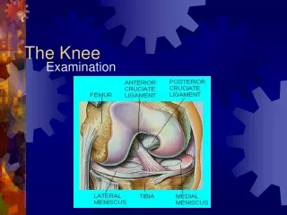
The Medial Support System of the Knee. Stephanie D Casey. The Knee. Three articulations The bones are connected together by the following ligaments: The Articular Capsule The Anterior Cruciate The Ligamentum Patellæ The Posterior Cruciate The Oblique Popliteal

E N D
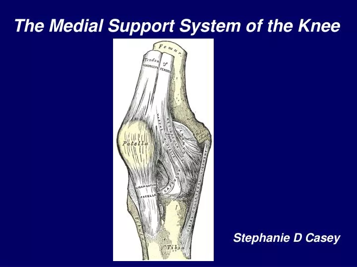
The Medial Support System of the Knee Stephanie D Casey
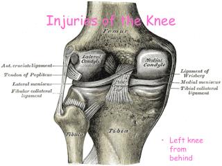
The Knee • Three articulations • The bones are connected together by the following ligaments: • The Articular Capsule • The Anterior Cruciate • The Ligamentum Patellæ • The Posterior Cruciate • The Oblique Popliteal • The Medial and Lateral Menisci • The Tibial Collateral • The Transverse • The Fibular Collateral • The Coronary.
The Origin of the Lecture Topic MINI PATHRIA
Overview • MEDIAL CAPSULOLIGAMENTOUS COMPLEX • Anatomy • Surgical • MRI • MEDIAL STABILIZERS OF THE PATELLA • Medial retinacular complex (MRC) • Anatomy • Surgical • MRI • Pathology • LAGNIAPPE
Medial Support System of the Knee The Big Picture
The layer approach presented here emphasizes anatomical relationships which have been established The layer concept is stressed because the ligaments of the medial side of the knee are condensations within tissue planes and not discrete structures such as the anterior cruciate ligament Any description of a ligament should specify its location within a plane, otherwise its relationship to the other structures will be confusing In order to locate all structures in their appropriate planes, it is helpful to think in terms of the three layers and the patterns by which they merge anteriorly and posteriorly
The Supporting Structures and Layers on theMedial Side of the Knee SUPERIOR MID INFERIOR Warren J. Bone Joint Surg. Am. 61:56-62, 1979
Surgical Anatomy Warren J. Bone Joint Surg. Am. 61:56-62, 1979
Layer I • Deep or crural fascia – invests satorius fascia • between the patella and its tendon anteriorly and the mid line of the popliteal fossa posteriorly • Proximally – continuous with the fascia overlying the quadriceps muscle • Posteriorly – deep fascia of lower extremity and can be traced upward and downward • Inferiorly – joins periosteum of tibia at tibial insertion of satorius tendon
MEDIAL CAPSULOLIGAMENTOUS COMPLEX CRANIAL TO JOINT LINE • LAYER I • Anterior to superficial medial ligament • Layer I joined Layer II to contribute to form parapatellar retinacular fibers
MEDIAL CAPSULOLIGAMENTOUS COMPLEXJOINT LINE • Layer I • Split in Layer II – fibers leaving Layer II and the superficial medial ligament to join layer I
MEDIAL CAPSULOLIGAMENTOUS COMPLEXCAUDAL TO JOINT LINE • Layer I • Layer I and II separated by ST and Gracilis tendons • Distally joins periosteum of tibia at satorius insertion
Layer 1Cranial to Joint Line Layer 1
Cranial to joint line VMO S
Caudal to Joint Line Layer II G Layer I ST
LAYER IIContents • Superficial Medial Ligament • Longitudinal (parallel) fibers • Oblique fibers • Vertical Split • Anterior to Split – Layer I + Layer II (LF) • Posterior to Split – Layer II (OF) + Layer III + Tendon Sheath of Semimenbranosus
Layer II - Levels • Proximal – Medial femoral condyle • Posterior to split – • From femoral condyle – transverse fibers form MPFL • Mid – vertical split • Anterior to split – cephalic extension of longitudinal fibers to vastus medialis to join layer 1 and form parapatellar retinaculum • PMC - Oblique fibers merge with Layer III • Semimembranosus sheath and its extensions • Distal – Tibia
MEDIAL CAPSULOLIGAMENTOUS COMPLEXCRANIAL TO JOINT LINE • LAYER II • Anterior to superficial medial ligament Layer I joined Layer II to contribute to form parapatellar retinacular fibers • Posterior • Layer II (POF) join Layer III + sheath of SMT
MEDIAL CAPSULOLIGAMENTOUS COMPLEXJOINT LINE • Layer II • Split in Layer II – fibers leaving Layer II and the superficial medial ligament to join layer I • Posterior Oblique Ligament • Layer II (POF) join Layer III + sheath of SMT
MEDIAL CAPSULOLIGAMENTOUS COMPLEXCAUDAL TO JOINT LINE • Layer II • Tibial insertion 5 cm below joint line
Cranial to Joint Line Layer II Posterior to the split Superficial MCL Fibers Cephalic Extension Transverse fibers MPFL
Cranial to Joint Line Layer I+II Layer III Layer II Layer I
Joint Line Longitudinal Fibers Superficial MCL Posterior Oblique Fibers
Joint Line Split in Layer II De Maeseneer RadioGraphics 2000; 20:S83–S89
Layer I + II Split in Layer II Layer II + III S SM G ST Layer I
Caudal to joint line Layer I Layer II SM Tendon
Layer III Contents • Capsule of the Knee joint • Proximal extent – follows contour of suprapatellar pouch and articular cartilage • Distal extent – meniscotibial ligament and articular cartilage • Anterior: capsule and patellomeniscal ligament • Mid: Deep fibers of MCL Meniscofemoral ligament Meniscotibial ligament • Posterior: Posteromedial capsule
MEDIAL CAPSULOLIGAMENTOUS COMPLEXCRANIAL TO JOINT LINE • LAYER III • Capsule • Meniscofemoral ligament • Posterior • Layer II (POF) join Layer III + sheath of SMT
MEDIAL CAPSULOLIGAMENTOUS COMPLEXJOINT LINE • Layer III-Capsule • Meniscotibial Ligament • Posterior Oblique Ligament • Layer II (POF) join Layer III + sheath of SMT
Joint Line Longitudinal Fibers Oblique Fibers Layer II + III SMT Sheath and POL
Joint Line Layer II+III SMT Sheath POL Layer I
Layer I+II Layer III Layer II Layer II Oblique Fibers POL SMT
Layer 1Cranial to Joint Line Crural fascia Layer I Layer II Layer III Satorius
Posteromedial Corner • Semimembranosus Tendon • 5 arms • Main insertion on the posteromedial tibial plateau • Oblique popliteal ligament • Semimenbranosus Tendon sheath • Fibers to Layer II • Superficial MCL fibers • Posterior oblique ligament • Capsule
Rectus femoris m. Vastus lateralis m. Vastus Medialis m. EXTENSOR MECHANISM OF THE KNEE QT Patella Medial patellar retinaculum Lateral patellar retinaculum Pes anserine tendons PT
CONDENSATION OF FIBERS IN THESE RESPECTIVE TISSUE PLANES
The Supporting Structures and Layers on theMedial Side of the Knee MEDIAL SOFT TISSUE RESTRAINTS OF THE EXTENSOR MECHANISM SUPERIOR MID INFERIOR Warren J. Bone Joint Surg. Am. 61:56-62, 1979 Conlan T etal, J. Bone Joint Surg. Am. 75:682-693, 1993
Patella Retinacula • The retinacula represent condensations in tissue planes rather than discrete structures • In the past, the description of these structures has been confusing in part because of their anatomic complexity and also because of variability in descriptive terminology. • Although descriptions of retinacular anatonly related to dissection can be found in the orthopedic literature similar descriptions in the radiologic literature have been oversimplified and limited to brief anatomic statements
3 Layers 1 - Superficial 2 – Intermediate 3 - Deep 3 Levels Superior Mid Inferior Medial Stabilizers of the Patella MERGE 4 DISTINCT ANATOMIC STRUCTURES MEDIAL PATELLOFEMORAL LIGAMENT MEDIAL PARAPATELLAR RETINACULUM MEDIAL PATELLOTIBIAL LIGAMENT MEDIAL PATELLOMENISCAL LIGAMENT SEPARATE
Most superficial Just deep to subcuetaneous tissues Deep crural fascia Anterosuperiorly – continuous with fascia overlying the VMO Layer 1 :Main componentligaments of medial retinacular complex
LAYER 1 VM S MSR
Layer 2 : • Fibers form inverted triangle • Central split in triangle defines 3 separate ligaments
Layer 2 :Main component ligaments of medial retinacular complex Medial patellofemoral ligament Medial patellotibial ligament Superficial MCL Medial Retinaculum – Parapatellar retinaculum Anterior to Superficial MCL Layer 2 (longitudinal fibers) + Layer 1
Layer 3 :Main componentligaments of medial retinacular complex • Joint capsule • Medial Patellomeniscal Ligament
Knowledge of the expected anatomic location of these four ligaments as well as their relationships to one another is crucial if one is to differentiate between them on MRI and thereby predict with accuracy which structures have been injured.
SUPERIOR VMO MPFL Main componentligaments of medial retinacular complex LAYER 2 MPFL LAYER 1 MERGES WITH VMO FASCIA VMO LAYER 1